. 2021 Jan 12;11(1):623.
doi: 10.1038/s41598-020-79845-3.

Contralesional functional network reorganization of the insular cortex in diffuse low-grade glioma patients

Affiliations

Contralesional functional network reorganization of the insular cortex in diffuse low-grade glioma patients

Shengyu Fang et al. Sci Rep. .

Abstract

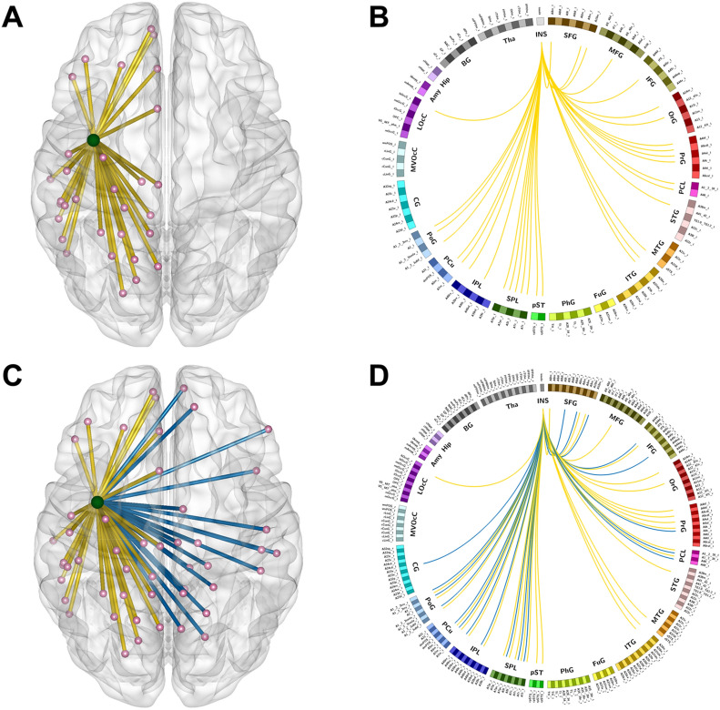
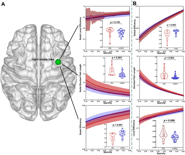
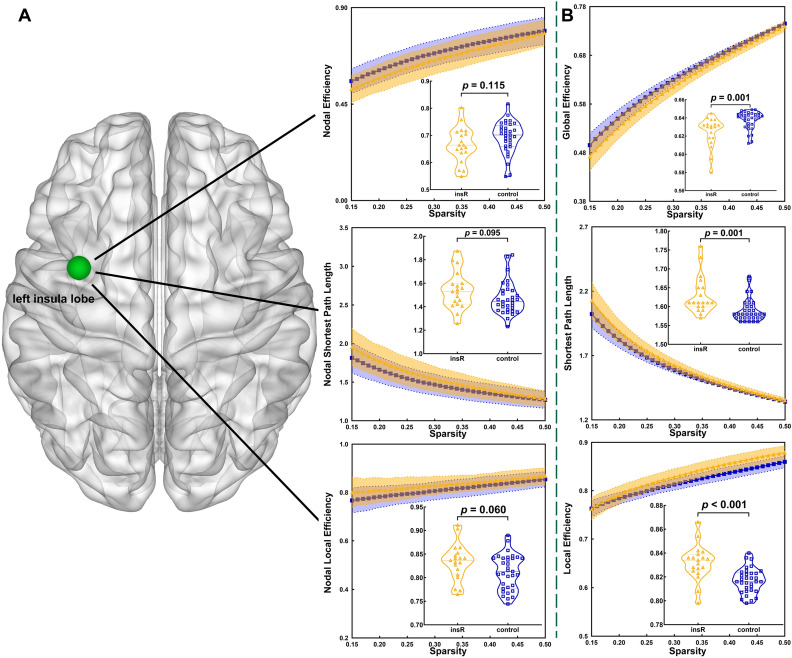
Diffuse low-grade gliomas (DLGGs) growing on the insular lobe induce contralesional hemispheric insular lobe compensation of damaged functioning by increasing cortical volumes. However, it remains unclear how functional networks are altered in patients with insular lobe DLGGs during functional compensation. Thirty-five patients with insular DLGGs were classified into the left (insL, n = 16) and right groups (insR, n = 19), and 33 healthy subjects were included in the control group. Resting state functional magnetic resonance imaging was used to generate functional connectivity (FC), and network topological properties were evaluated using graph theoretical analysis based on FC matrices. Network-based statistics were applied to compare differences in the FC matrices. A false discovery rate was applied to correct the topological properties. There was no difference in the FC of edges between the control and insL groups; however, the nodal shortest path length of the right insular lobe was significantly increased in the insL group compared to the control group. Additionally, FC was increased in the functional edges originating from the left insular lobe in the insR group compared to the control group. Moreover, there were no differences in topological properties between the insR and control groups. The contralesional insular lobe is crucial for network alterations. The detailed patterns of network alterations were different depending on the affected hemisphere. The observed network alterations might be associated with functional network reorganization and functional compensation.


Conflict of interest statement

The authors declare no competing interests.

Figures

Figure 1
Figure 1
In both, healthy hemisphere and whole brain template, altered functional edges originating from the contralateral insular lobe and functional connectivity in the insR groups were observed compared to those in the healthy group. (A,B) Altered functional edges basing on the healthy hemispheric template. (C,D) Altered functional edges basing on the whole brain template. Light green node = left insular lobe. The yellow lines present the edges connected left insula to other parts in left hemisphere and blue lines present the edges connected left insula to other parts in right hemisphere.
Figure 2
Figure 2
In the whole brain template, compared to the healthy group, altered topological properties were observed in the insL group. (A) Altered topological properties at the local level of the insular lobe node. (B) Altered topological properties at the global level. Light green node = right insular lobe.
Figure 3
Figure 3
In the whole brain template, compared to the healthy group, altered topological properties were observed in the insR group. (A) Altered topological properties at the local level of the insular lobe node. (B) Altered topological properties at the global level. Light green node = left insular lobe.

Similar articles

Cited by

References

    1. Duffau H. The huge plastic potential of adult brain and the role of connectomics: New insights provided by serial mappings in glioma surgery. Cortex. 2014;58:325–337. doi: 10.1016/j.cortex.2013.08.005. - DOI - PubMed
    1. Heiss WD, Thiel A, Kessler J, Herholz K. Disturbance and recovery of language function: Correlates in PET activation studies. Neuroimage. 2003;20(Suppl 1):S42–49. doi: 10.1016/j.neuroimage.2003.09.005. - DOI - PubMed
    1. Darainy M, Vahdat S, Ostry DJ. Neural basis of sensorimotor plasticity in speech motor adaptation. Cereb. Cortex. 2018 doi: 10.1093/cercor/bhy153. - DOI - PMC - PubMed
    1. Gunal V, Savardekar AR, Devi BI, Bharath RD. Preoperative functional magnetic resonance imaging in patients undergoing surgery for tumors around left (dominant) inferior frontal gyrus region. Surg. Neurol. Int. 2018;9:126. doi: 10.4103/sni.sni_414_17. - DOI - PMC - PubMed
    1. Crossley NA, et al. The hubs of the human connectome are generally implicated in the anatomy of brain disorders. Brain. 2014;137:2382–2395. doi: 10.1093/brain/awu132. - DOI - PMC - PubMed

Publication types