. 2021 Jan 12;11(1):702.
doi: 10.1038/s41598-020-80308-y.

A novel retinal ganglion cell quantification tool based on deep learning

Affiliations

A novel retinal ganglion cell quantification tool based on deep learning

Luca Masin et al. Sci Rep. .

Abstract

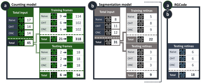
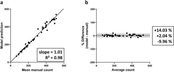
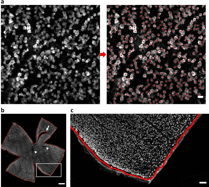
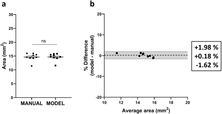
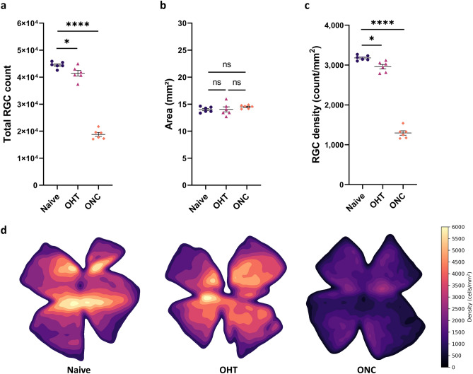
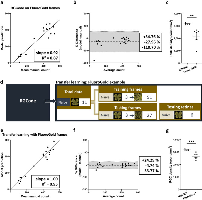
Glaucoma is a disease associated with the loss of retinal ganglion cells (RGCs), and remains one of the primary causes of blindness worldwide. Major research efforts are presently directed towards the understanding of disease pathogenesis and the development of new therapies, with the help of rodent models as an important preclinical research tool. The ultimate goal is reaching neuroprotection of the RGCs, which requires a tool to reliably quantify RGC survival. Hence, we demonstrate a novel deep learning pipeline that enables fully automated RGC quantification in the entire murine retina. This software, called RGCode (Retinal Ganglion Cell quantification based On DEep learning), provides a user-friendly interface that requires the input of RBPMS-immunostained flatmounts and returns the total RGC count, retinal area and density, together with output images showing the computed counts and isodensity maps. The counting model was trained on RBPMS-stained healthy and glaucomatous retinas, obtained from mice subjected to microbead-induced ocular hypertension and optic nerve crush injury paradigms. RGCode demonstrates excellent performance in RGC quantification as compared to manual counts. Furthermore, we convincingly show that RGCode has potential for wider application, by retraining the model with a minimal set of training data to count FluoroGold-traced RGCs.


Conflict of interest statement

The authors declare no competing interests.

Figures

Figure 1
Figure 1
Overview of the datasets used to develop RGCode. Composition of the training and testing datasets for both the counting (a) and segmentation (b) models are depicted. Frames were sampled from different retinal areas and experimental conditions (naive, OHT- and ONC-injured retinas). Additionally, 18 entire flatmounts were reserved for evaluating the performance of the complete pipeline, comprising both the counting and segmentation algorithms (a+b). Key: Naive, uninjured retinas; OHT, 5 weeks post microbead-induced ocular hypertension retinas; ONC, 7 days post optic nerve crush injury retinas.
Figure 2
Figure 2
Linear regression and bias analysis of automated counts on retinal frames. (a) Linear regression analysis for the average of the manual counts versus model output indicates that the automated counts show a very high linear relationship with manual counts. Best-fit linear regression correlation coefficient (R2 and slope) are indicated. (b) Bland–Altman analysis of the automated counts compared to the operator average, showing mean bias ±95% limits of agreement.
Figure 3
Figure 3
Representative examples of the output of RGCode on testing data. (a) Testing frame sampled from the mid-peripheral region of a naive retina, showing the input and output of RGCode. In general, RGCode consistently detects and counts RBPMS+ cells and clearly distinguishes overlapping cells. Scale bar =20μm. (b, c) Naive testing retina showing the output of the segmentation model. Scale bars = 500 μm and 100μm (zoom).
Figure 4
Figure 4
Estimation of retinal area and bias analysis of automated segmentation on retinal flatmounts. (a) Comparison of manual and automated segmentation of the retinal area (mean ± SEM) revealing no significant difference between both methods (unpaired two-tailed t test). (b) Bland–Altman analysis of the automated versus manual segmentation. Mean bias ± 95% limits of agreement are shown.
Figure 5
Figure 5
Outcome of RGCode on testing retinas: automated RGC count, retinal segmentation, RGC density and isodensity maps across all conditions (Naive, purple dots; OHT, pink triangles and ONC, orange squares). (a) A significant decrease in RGC count is seen in glaucomatous conditions as opposed to uninjured retinas (one-way ANOVA with Tukey’s post-hoc test, p=0.0372, p<0.0001). (b) The total area does not significantly differ between uninjured and injured retinas (one-way ANOVA with Tukey’s post-hoc test). (c) RGC density is significantly lower when comparing glaucomatous versus uninjured retinas (one-way ANOVA with Tukey’s post-hoc test, p=0.0145, p<0.0001). Data are depicted as mean ± SEM. (d) Representative pseudo-colour representations ranging from 0 RGCs/mm2 (black tone) to 6000 RGCs/mm2 (yellow tone) of uninjured and injured (OHT and ONC) retinas. A central-to-peripheral gradient in RGC density can be observed in the uninjured retina. Modest (OHT) and substantial (ONC) RGC loss is detected in the glaucomatous retinas. Key: Naive, uninjured retinas; OHT, 5 weeks post microbead-induced ocular hypertension retinas; ONC, 7 days post optic nerve crush injury retinas.
Figure 6
Figure 6
Transfer learning of RGCode for Fluorogold labelling. (a, b) Linear regression and Bland Altman analysis (mean bias ± 95% limits of agreement) after running RGCode on Fluorogold-labelled RGCs. Counting performance was considerably lower as compared to the RBPMS dataset, whereas a higher bias was observed. (c) the lower performance of RGCode on FluoroGold-traced flatmounts resulted in a high variability in density (mean ± SEM, unpaired, two-tailed t test, **p = 0.0042; ***p = 0.0007). (d) Composition of the training and testing dataset used for transfer learning. (e,f) Transfer learning of RGCode with a minimal set of new training data reveals a high accuracy with linear regression and Bland–Altman analysis. Mean bias ± 95% limits of agreement are depicted. (g) Transfer learning results in a lower variability in the RGC density measurements. As expected, the density of FluoroGold+ RGCs is significantly lower as compared to RBPMS+ ones (unpaired, two-tailed t test, ***p = 0.007).

Similar articles

Cited by

References

    1. Mead B, Tomarev S. Evaluating retinal ganglion cell loss and dysfunction. Exp. Eye Res. 2016;151:96–106. doi: 10.1016/j.exer.2016.08.006. - DOI - PMC - PubMed
    1. Guo L, Cordeiro MF. Assessment of neuroprotection in the retina with DARC. Prog. Brain Res. 2008;173:437–450. doi: 10.1016/S0079-6123(08)01130-8. - DOI - PMC - PubMed
    1. Köbbert C, et al. Current concepts in neuroanatomical tracing. Prog. Neurobiol. 2000;62:327–351. doi: 10.1016/S0301-0082(00)00019-8. - DOI - PubMed
    1. Abdel-Majid RM, Archibald ML, Tremblay F, Baldridge WH. Tracer coupling of neurons in the rat retina inner nuclear layer labeled by Fluorogold. Brain Res. 2005;1063:114–120. doi: 10.1016/j.brainres.2005.09.046. - DOI - PubMed
    1. Peinado-Ramon P, Salvador M, Villegas-Perez MP, Vidal-Sanz M. Effects of axotomy and intraocular administration of NT-4, NT-3, and brain-derived neurotrophic factor on the survival of adult rat retinal ganglion cells. A quantitative in vivo study. Invest. Ophthalmol. Vis. Sci. 1996;37:489–500. - PubMed

Publication types