. 2021 Jan 12;11(1):205.
doi: 10.1038/s41598-020-80316-y.

Intramedullary injury combined with osteoporosis therapeutics regulates targeted local osteogenesis

Affiliations

Intramedullary injury combined with osteoporosis therapeutics regulates targeted local osteogenesis

Yoko Miyazaki-Asato et al. Sci Rep. .

Abstract

Bone marrow ablation prompts transient bone formation in nearly the entire medullary cavity before marrow regeneration occurs. Here, we establish a procedure to direct bone formation in a desired particular site within the medullary cavity for support of biomedical devices. Local intramedullary injury was performed in the tibiae of rats and parathyroid hormone (PTH), alendronate, or saline was administered. Newly generated bone in the medulla was assessed by micro-CT and histology. To evaluate the function of newly generated bone, animals received intramedullary injury in tibiae followed by daily PTH. At day-14, implants were placed in the endocortical bone and the bone response to the implants was assessed. The fate of newly generated bone was compared with and without implants. We found that neither intramedullary injury nor medication alone resulted in bone formation. However, when combined, substantial bone was generated locally inside the diaphyseal medulla. Newly formed bone disappeared without implant placement but was retained with implants. Bone was especially retained around and between the implants. This study found that local bone marrow disruption followed by PTH or alendronate generated substantial cancellous bone locally in the diaphyseal medulla. This approach offers promise as a tissue engineering tool in medicine and dentistry.


Conflict of interest statement

The authors declare no competing interests.

Figures
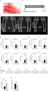
Figure 1
Figure 1
PTH and ALN administrations increased bone mass in rats with substantial trabecular bone formation at the intramedullary injury site. (a) An intramedullary injury was created in the diaphysis of the left tibia and sham surgery was performed in the right tibia (n = 33). (b) Eleven rats received 20 µg/kg PTH daily; another 11 rats received 0.4 mg/kg ALN twice a week; the remaining 11 rats received VC (saline) for 14 days. (c) Representative micro-CT images of tibiae. The trabecular bone within the rectangular boxes in the metaphysis was assessed in the sham tibia to evaluate the skeletal effect of medications. Arrowheads indicate the intramedullary injury sites. Drill holes are not completely repaired in the VC group. Considerable endocortical ossified tissue was noted at the intramedullary injury sites in the PTH and ALN groups. Newly generated bone in the medullary cavity was exclusively found in the intramedullary disturbed tibiae. Minimal trabecular bone was observed in the diaphyseal medullary cavity of the sham tibiae. Since sham sites had very little to no bone to quantify regardless of treatment, the trabecular bone at the local intramedullary injury site was the focus of quantitative analysis. In the metaphysis of the sham tibia, PTH and ALN administration effects were measured via micro-CT analysis showing (d) bone volume fraction (BV/TV), (e) trabecular thickness (Tb.Th), (f) trabecular number (Tb.N), (g) trabecular separation (Tb.Sp) and (h) tissue mineral density (TMD). In the diaphysis of the intramedullary injured tibia, micro-CT analysis was performed and graphs show (i) BV/TV, (j) Tb.Th, (k) Tb.N, (l) Tb.Sp and (m) TMD. (n) A paired t-test was performed to compare bone parameters between the metaphyseal trabecular bone and the newly generated bone in the diaphysis. p values are shown. Connectivity density (o) and the degree of anisotropy (p) of the diaphyseal trabecular bone was compared between the PTH and ALN groups. All micro-CT data were processed using CTAn software (version 1.16.4.1, http://bruker-microct.com/products/downloads.htm#ctan) which was provided with the purchase of the Bruker X-ray micro-CT system. **p < 0.01; ***p < 0.001 vs. VC, #p < 0.05; ##p < 0.01 between PTH and ALN.
Figure 2
Figure 2
ALN induced greater diaphyseal trabecular bone with suppressed osteoclasts than VC or PTH. (a) Representative images of HE-stained sections of tibiae at the injury level. Arrows indicate drilling sites; open circles indicate newly formed ossified tissue; closed circles indicate periosteal ossified tissue. (b) The average endosteal bone area at the local intramedullary injury level was significantly larger in the PTH and ALN groups than the VC group. No significant difference was found between the PTH and ALN groups although a trend of the larger bone area in the ALN vs. PTH group was observed. (c) Representative images of TRAP-stained sections of the endocortical ossified tissue. (d) Osteoclast numbers per bone perimeter were significantly smaller in the ALN group than the VC or PTH group. No differences were noted between the PTH and VC groups. *p < 0.05 vs. VC, #p < 0.05 between PTH and ALN.
Figure 3
Figure 3
Serum biochemistry. (a) Serum calcium levels were maintained within the normal range regardless of treatment. (b) No statistical differences were noted in the serum TRAcP5b levels in all treatment groups relative to BL. There was a significant difference at day-14 between the PTH and ALN groups. (c) Serum osteocalcin levels were significantly elevated in the PTH group at day-14 in comparison to BL and the other groups at day-14. No difference was noted between the BL and day-14 ALN or VC groups. **p < 0.01 vs. BL, #p < 0.05 PTH vs. ALN.
Figure 4
Figure 4
Implant placement contributed to the survival of the diaphyseal trabecular bone. (a) Seven rats received intramedullary injury in both tibiae, followed by PTH administration for 14 days. After the completion of treatment, screw implants were placed in the left tibia and sham surgery was performed on the right tibia. Rats were euthanized 3 weeks after implant placement and survival of trabecular bone in the medullary cavity was assessed. (b) Representative images of HE-stained sections and (c) micro-CT images of tibiae at the injury level. A significant amount of trabecular bone was retained in the medullary cavity at implant sites, while minimum trabecular bone was observed in sham sites. Considerable periosteal bone was retained in both implant and sham sites. (d) The cartoon indicates the bone segments assessed. Micro-CT analysis results depicted in graphs showing the bone volume fraction (e) was higher in the bone around implants than the bone at the mesial and distal ends. (f) Trabecular thickness was larger in the bone around implants than in the bone between implants or at the mesial and distal ends. The endocortical trabecular number (g) and separation (h) were similar between the bone segments. All micro-CT data were processed using CTAn software (version 1.16.4.1, http://bruker-microct.com/products/downloads.htm#ctan) which was provided with the purchase of the Bruker X-ray micro-CT system. *: p < 0.05, ***: p < 0.01.

Similar articles

References

    1. Justesen J, et al. Adipocyte tissue volume in bone marrow is increased with aging and in patients with osteoporosis. Biogerontology. 2001;2:165–171. doi: 10.1023/a:1011513223894. - DOI - PubMed
    1. Jilka RL. Molecular and cellular mechanisms of the anabolic effect of intermittent PTH. Bone. 2007;40:1434–1446. doi: 10.1016/j.bone.2007.03.017. - DOI - PMC - PubMed
    1. Rogers MJ, Crockett JC, Coxon FP, Monkkonen J. Biochemical and molecular mechanisms of action of bisphosphonates. Bone. 2011;49:34–41. doi: 10.1016/j.bone.2010.11.008. - DOI - PubMed
    1. Larsson L, et al. Regenerative medicine for periodontal and peri-implant diseases. J. Dent. Res. 2016;95:255–266. doi: 10.1177/0022034515618887. - DOI - PMC - PubMed
    1. Hailer NP, Garellick G, Karrholm J. Uncemented and cemented primary total hip arthroplasty in the Swedish Hip Arthroplasty Register. Acta Orthop. 2010;81:34–41. doi: 10.3109/17453671003685400. - DOI - PMC - PubMed

MeSH terms