. 2021 Jan 12;11(1):753.
doi: 10.1038/s41598-020-80258-5.

The N-terminal region of Jaw1 has a role to inhibit the formation of organized smooth endoplasmic reticulum as an intrinsically disordered region

Affiliations

The N-terminal region of Jaw1 has a role to inhibit the formation of organized smooth endoplasmic reticulum as an intrinsically disordered region

Takuma Kozono et al. Sci Rep. .

Abstract

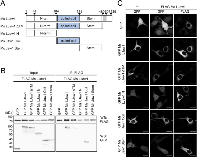
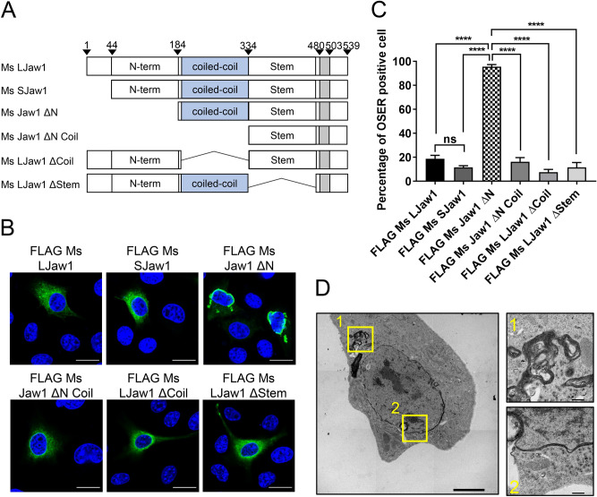
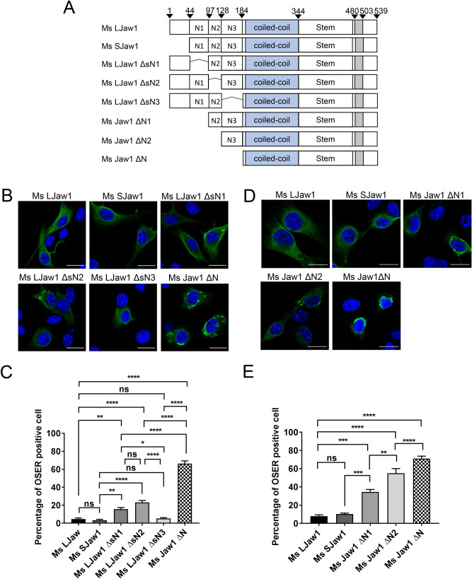
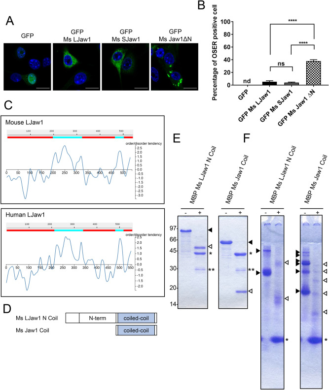
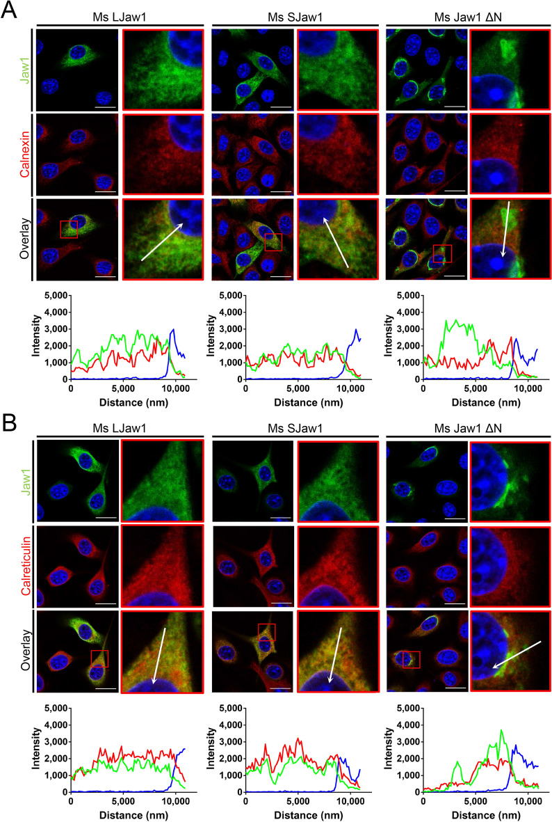
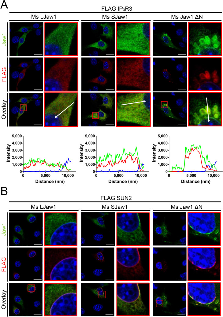
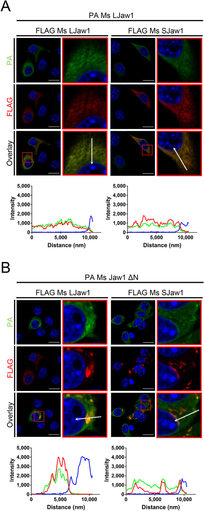
Jaw1/LRMP is a type II integral membrane protein that is localized at the endoplasmic reticulum (ER) and outer nuclear membrane. We previously reported that a function of Jaw1 is to maintain the nuclear shape as a KASH protein via its carboxyl terminal region, a component of linker of nucleoskeleton and cytoskeleton complex in the oligomeric state. Although the oligomerization of some KASH proteins via the cytosolic regions serves to stabilize protein-protein interactions, the issue of how the oligomerization of Jaw1 is regulated is not completely understood. Therefore, we focused on three distinct regions on the cytosolic face of Jaw1: the N-terminal region, the coiled-coil domain and the stem region, in terms of oligomerization. A co-immunoprecipitation assay showed that its coiled-coil domain is a candidate for the oligomerization site. Furthermore, our data indicated that the N-terminal region prevents the aberrant oligomerization of Jaw1 as an intrinsically disordered region (IDR). Importantly, the ectopic expression of an N-terminal region deleted mutant caused the formation of organized smooth ER (OSER), structures such as nuclear karmellae and whorls, in B16F10 cells. Furthermore, this OSER interfered with the localization of the oligomer and interactors such as the type III inositol 1,4,5-triphosphate receptor (IP3R3) and SUN2. In summary, the N-terminal region of Jaw1 inhibits the formation of OSER as an IDR to maintain the homeostatic localization of interactors on the ER membrane.


Conflict of interest statement

The authors declare no competing interests.

Figures

Figure 1
Figure 1
Identification of the cytosolic oligomerization site. (A) Schematic representation of Mouse LJaw1 (LJaw1; long form Jaw1) and mutants. N-term; N-terminal region, coiled-coil; coiled-coil domain, Stem; stem region, Gray box; single trans-membrane domain. (B) Identification of the oligomerization site by co-immunoprecipitation followed by western blotting. FLAG Ms LJaw1 was co-expressed with GFP, GFP Ms LJaw1 or mutants in HEK293 cells by transfection. After incubation for 24 h, the lysates were subjected to co-immunoprecipitation. For western blotting, an anti-FLAG rabbit antibody and an anti-GFP rabbit antibody as primary antibodies were used. (C) GFP, GFP Ms LJaw1 or mutants was expressed alone or co-expressed with FLAG Ms LJaw1 in B16F10 cells by transfection. After incubation for 24 h, the cells were subjected to immunostaining using an anti-FLAG rabbit antibody as a primary antibody. The images were acquired by confocal microscopy. Scale bar; 20 μm.
Figure 2
Figure 2
Observation of the OSER by the expression of Jaw1 ΔN. (A) Schematic representation of Mouse Jaw1 (LJaw1; long form Jaw1, SJaw1; short form Jaw1) and mutants lacking the N-terminal region (Ms Jaw1 ΔN), N-terminal region and coiled-coil domain (Ms Jaw1 ΔN Coil), coiled-coil domain (Ms LJaw1 ΔCoil) and stem region (Ms LJaw1 ΔStem). (B) FLAG Ms LJaw1, FLAG Ms SJaw1 or deletion mutants were expressed in B16F10 cells by transfection. After incubation for 24 h, immunostaining was performed using an anti-FLAG rabbit antibody (green). Nuclei were stained with Hoechst33342 (blue). The images were acquired by confocal microscopy. Scale bar; 20 μm. (C) Counting of the cells having OSER structures out of the FLAG positive cells in (B) (n = 100). In the graph, the percentage of cells with OSER structures is shown based on the average of four independent experiments per condition. Error bars show the S. D. “ns”, not significant; ****P < 0.0001, Turkey Kramer’s t-test. (D) Electron micrographs of the B16F10 cells expressing FLAG Ms Jaw1 ΔN. The magnified images are shown in the boxes at right (box 1; whorls and cisternae stacks, box 2; nuclear karmellae). Scale bar; left box; 5.0 μm, right boxes; 500 nm. The OSER positive cells were classified as the ones with a highly dense membranous structure (N = 8).
Figure 3
Figure 3
Exploration of the specific N-terminal region involved with the inhibition of OSER formation. (A) Schematic representation of Mouse Jaw1s or deletion mutants lacking the limited N-terminal region (Ms LJaw1 ΔsN1, Ms LJaw1 ΔsN2 and Ms LJaw1 ΔsN3) and deletion mutants with N-terminal regions of different lengths (Ms Jaw1 ΔN1, Ms Jaw1 ΔN2 and Ms Jaw1 ΔN). (B,D) Ms LJaw1, Ms SJaw1, Ms LJaw1 ΔsN1, Ms LJaw1 ΔsN2, Ms LJaw1 ΔsN3 or Ms Jaw1 ΔN (B) and Ms LJaw1, Ms SJaw1, Ms Jaw1 ΔN1, Ms Jaw1 ΔN2 or Ms Jaw1 ΔN (D) were expressed in B16F10 cells by transfection. After incubation for 24 h, immunostaining was performed using an anti-Jaw1 rat antibody (green). Nuclei were stained with Hoechst33342 (blue). The images were acquired by confocal microscopy. Scale bar; 20 μm. (C,E) Counting of the cells having OSER structures out of the Jaw1 positive cells in (B,D) (n = 100). In the graph, the percentage of cells with OSER structures is shown based on the average of four independent experiments per condition. Error bars show the S. D. “ns”, not significant; *P < 0.05; **P < 0.01; ***P < 0.001; ****P < 0.0001, Turkey Kramer’s t-test.
Figure 4
Figure 4
Investigation directed to which Jaw1 N-terminal region inhibits OSER formation. (A) GFP, GFP Ms LJaw1, GFP Ms SJaw1 or GFP Ms Jaw1 ΔN were expressed in B16F10 cells by transfection. After incubation for 24 h, the cells were fixed and incubated with PBS containing Hoechst33342 (blue). The images were acquired by confocal microscopy. Scale bar; 20 μm. (B) Counting of cells having OSER structures out of the GFP positive cells in (A) (n = 100). In the graph, the percentage of cells with OSER structures is shown based on the average of four independent experiments per condition. Error bars show the S. D. Not detectable is represented as “nd”. “ns”, not significant; ****P < 0.0001, Turkey Kramer’s t-test. (C) DICHOT analysis to search the ordered or disordered region within Jaw1. Entire amino acids sequences of Ms LJaw1 (upper) and Hu LJaw1 (bottom) were subjected to analysis by the DICHOT system. Colored areas show ordered (blue) and disordered (red) regions. The number in the side axis shows the position of the amino acids. (D) Schematic representation of the mutants bearing N-terminal region and coiled-coil domain (Ms LJaw1 N Coil) and coiled-coil domain alone (Ms Jaw1 Coil). (E,F) MBP Ms LJaw1 N Coil or MBP Ms Jaw1 Coil were expressed in E. coli and purified using amylose resin. The recombinant proteins treated with/without TEV protease to remove MBP tag were subjected to SDS-PAGE (E) and Native-PAGE (F) followed by CBB staining. Closed triangles, the bands of MBP tagged Jaw1; opened triangles, the bands of MBP removed Jaw1; *the band of cleaved MBP tag; **the band of TEV protease; minus, no treatment with TEV protease; plus, treatment with TEV protease.
Figure 5
Figure 5
Evaluation of ER morphology under OSER formation. (A,B) Ms LJaw1, Ms SJaw1 or Ms Jaw1 ΔN were expressed in B16F10 cells by transfection. After incubation for 24 h, immunostaining was performed using an anti-Jaw1 rat antibody and an anti-calnexin rabbit antibody (A) or an anti-calreticulin rabbit antibody (B). Nuclei were stained with Hoechst33342. The images were acquired by confocal microscopy. Scale bar; 20 μm. The magnified images corresponding to the area surrounded with red lines in each image were generated and line plot profiles were performed along the arrows. Green; Jaw1, red; calnexin (A) or calreticulin (B), blue; Hoechst33342.
Figure 6
Figure 6
Evaluation of the effects under OSER on the localization of the interactors. A, (B) Ms LJaw1, Ms SJaw1 or Ms Jaw1 ΔN were co-expressed with FLAG IP3R3 (A) or FLAG SUN2 (B) in B16F10 cells by transfection. After incubation for 24 h, immunostaining was performed using an anti-Jaw1 rat antibody and an anti-FLAG rabbit antibody. Nuclei were stained with Hoechst33342. The images were acquired by confocal microscopy. Scale bar; 20 μm. The magnified images corresponding to the area surrounded with red lines in each image (A,B) were generated and line plot profiles were performed along the arrows (A). Green; Jaw1, red; FLAG, blue; Hoechst33342.
Figure 7
Figure 7
Evaluation of the effects under OSER on the localization of the oligomer. (A,B) FLAG Ms LJaw1 or FLAG Ms SJaw1 were co-expressed with PA Ms LJaw1 (A) or PA Ms Jaw1 ΔN (B) in B16F10 cells by transfection. After incubation for 24 h, immunostaining was performed using an anti-FLAG rabbit antibody and an anti-PA rat antibody. Nuclei were stained with Hoechst33342. The images were acquired by confocal microscopy. Scale bar; 20 μm. The magnified images corresponding to the area surrounded with red lines in each image were generated and line plot profiles were performed along the arrows. Green; PA, red; FLAG, blue; Hoechst 33342.

Similar articles

Cited by

References

    1. Zhang H, Hu J. Shaping the endoplasmic reticulum into a social network. Trends Cell Biol. 2016;26:934–943. doi: 10.1016/j.tcb.2016.06.002. - DOI - PubMed
    1. Klopfenstein DR, et al. Subdomain-specific localization of CLIMP-63 (p63) in the endoplasmic reticulum is mediated by its luminal alpha-helical segment. J. Cell Biol. 2001;153:1287–1300. doi: 10.1083/jcb.153.6.1287. - DOI - PMC - PubMed
    1. Shibata Y, et al. Mechanisms determining the morphology of the peripheral ER. Cell. 2010;143:774–788. doi: 10.1016/j.cell.2010.11.007. - DOI - PMC - PubMed
    1. Voeltz GK, Prinz WA, Shibata Y, Rist JM, Rapoport TA. A class of membrane proteins shaping the tubular endoplasmic reticulum. Cell. 2006;124:573–586. doi: 10.1016/j.cell.2005.11.047. - DOI - PubMed
    1. Starr DA. A nuclear envelope bridge positions nuclei and moves chromosomes. J. Cell Sci. 2009;122:577–586. doi: 10.1242/jcs.037622. - DOI - PMC - PubMed

Publication types

MeSH terms

Substances

LinkOut - more resources