

Evaluation of a multiparametric MRI radiomic-based approach for stratification of equivocal PI-RADS 3 and upgraded PI-RADS 4 prostatic lesions
- PMID: 33436929
- PMCID: PMC7804929
- DOI: 10.1038/s41598-020-80749-5
Evaluation of a multiparametric MRI radiomic-based approach for stratification of equivocal PI-RADS 3 and upgraded PI-RADS 4 prostatic lesions
Abstract
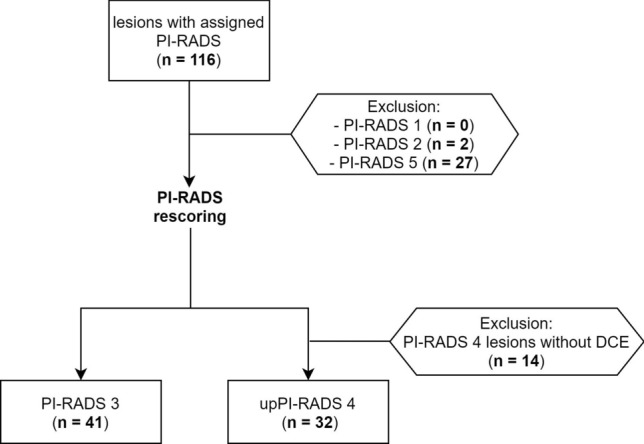
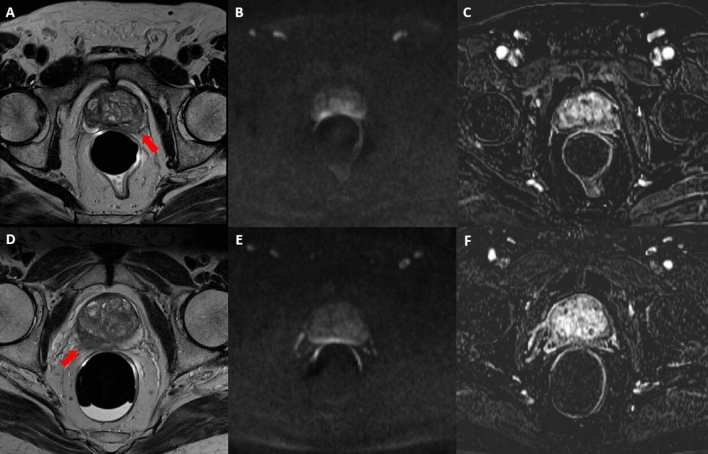
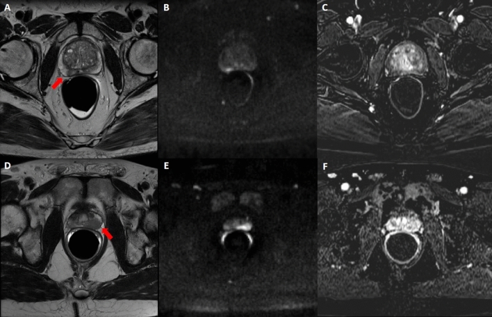
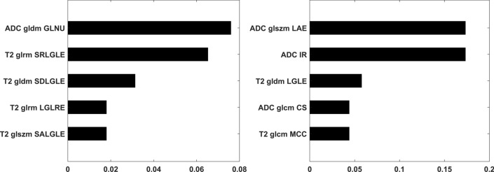
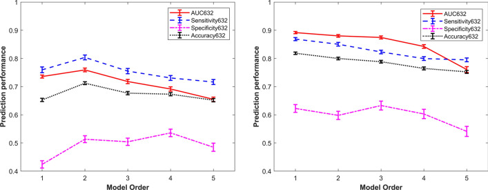
Despite the key-role of the Prostate Imaging and Reporting and Data System (PI-RADS) in the diagnosis and characterization of prostate cancer (PCa), this system remains to be affected by several limitations, primarily associated with the interpretation of equivocal PI-RADS 3 lesions and with the debated role of Dynamic Contrast Enhanced-Magnetic Resonance Imaging (DCE-MRI), which is only used to upgrade peripheral PI-RADS category 3 lesions to PI-RADS category 4 if enhancement is focal. We aimed at investigating the usefulness of radiomics for detection of PCa lesions (Gleason Score ≥ 6) in PI-RADS 3 lesions and in peripheral PI-RADS 3 upgraded to PI-RADS 4 lesions (upPI-RADS 4). Multiparametric MRI (mpMRI) data of patients who underwent prostatic mpMRI between April 2013 and September 2018 were retrospectively evaluated. Biopsy results were used as gold standard. PI-RADS 3 and PI-RADS 4 lesions were re-scored according to the PI-RADS v2.1 before and after DCE-MRI evaluation. Radiomic features were extracted from T2-weighted MRI (T2), Apparent diffusion Coefficient (ADC) map and DCE-MRI subtracted images using PyRadiomics. Feature selection was performed using Wilcoxon-ranksum test and Minimum Redundancy Maximum Relevance (mRMR). Predictive models were constructed for PCa detection in PI-RADS 3 and upPI-RADS 4 lesions using at each step an imbalance-adjusted bootstrap resampling (IABR) on 1000 samples. 41 PI-RADS 3 and 32 upPI-RADS 4 lesions were analyzed. Among 293 radiomic features, the top selected features derived from T2 and ADC. For PI-RADS 3 stratification, second order model showed higher performances (Area Under the Receiver Operating Characteristic Curve-AUC- = 80%), while for upPI-RADS 4 stratification, first order model showed higher performances respect to superior order models (AUC = 89%). Our results support the significant role of T2 and ADC radiomic features for PCa detection in lesions scored as PI-RADS 3 and upPI-RADS 4. Radiomics models showed high diagnostic efficacy in classify PI-RADS 3 and upPI-RADS 4 lesions, outperforming PI-RADS v2.1 performance.
Conflict of interest statement
The authors declare no competing interests.
Figures
Similar articles
-
Prostate Cancer Differentiation and Aggressiveness: Assessment With a Radiomic-Based Model vs. PI-RADS v2.J Magn Reson Imaging. 2019 Mar;49(3):875-884. doi: 10.1002/jmri.26243. Epub 2018 Sep 19. J Magn Reson Imaging. 2019. PMID: 30230108 Free PMC article.
-
Multiparametric MRI to Predict Gleason Score Upgrading and Downgrading at Radical Prostatectomy Compared to Presurgical Biopsy.Korean J Radiol. 2025 May;26(5):422-434. doi: 10.3348/kjr.2024.1008. Epub 2025 Mar 21. Korean J Radiol. 2025. PMID: 40169496 Free PMC article.
-
Comparison of PI-RADS v1 and v2 for multiparametric MRI detection of prostate cancer with whole-mount histological workup as reference standard.Eur J Radiol. 2019 Jul;116:180-185. doi: 10.1016/j.ejrad.2019.04.012. Epub 2019 May 14. Eur J Radiol. 2019. PMID: 31153562
-
Prostate imaging reporting and data system version 2 (PI-RADS v2): a pictorial review.Abdom Radiol (NY). 2017 Jan;42(1):278-289. doi: 10.1007/s00261-016-0871-z. Abdom Radiol (NY). 2017. PMID: 27522352 Free PMC article. Review.
-
Current Status of Biparametric MRI in Prostate Cancer Diagnosis: Literature Analysis.Life (Basel). 2022 May 28;12(6):804. doi: 10.3390/life12060804. Life (Basel). 2022. PMID: 35743835 Free PMC article. Review.
Cited by
-
Predicting prostate cancer in men with PSA levels of 4-10 ng/mL: MRI-based radiomics can help junior radiologists improve the diagnostic performance.Sci Rep. 2023 Mar 24;13(1):4846. doi: 10.1038/s41598-023-31869-1. Sci Rep. 2023. PMID: 36964192 Free PMC article.
-
MRI-derived radiomics models for diagnosis, aggressiveness, and prognosis evaluation in prostate cancer.J Zhejiang Univ Sci B. 2023 Aug 15;24(8):663-681. doi: 10.1631/jzus.B2200619. J Zhejiang Univ Sci B. 2023. PMID: 37551554 Free PMC article. Review.
-
Biparametric MRI-based radiomics for prediction of clinically significant prostate cancer of PI-RADS category 3 lesions.BMC Cancer. 2025 Apr 5;25(1):615. doi: 10.1186/s12885-025-14022-1. BMC Cancer. 2025. PMID: 40188349 Free PMC article.
-
Improving risk stratification of PI-RADS 3 + 1 lesions of the peripheral zone: expert lexicon of terms, multi-reader performance and contribution of artificial intelligence.Cancer Imaging. 2025 Aug 19;25(1):102. doi: 10.1186/s40644-025-00916-7. Cancer Imaging. 2025. PMID: 40830988 Free PMC article.
-
Magnetic resonance imaging radiomics-based prediction of clinically significant prostate cancer in equivocal PI-RADS 3 lesions in the transitional zone.Front Oncol. 2023 Nov 23;13:1247682. doi: 10.3389/fonc.2023.1247682. eCollection 2023. Front Oncol. 2023. PMID: 38074651 Free PMC article.
References
-
- Hegde JV, Mulkern RV, Panych LP, Fennessy FM, Fedorov A, Maier SE, Tempany CMC. Multiparametric MRI of prostate cancer: An update on state-of-the-art techniques and their performance in detecting and localizing prostate cancer. J. Magn. Reson. Imaging. 2013;37:1035–1054. doi: 10.1002/jmri.23860. - DOI - PMC - PubMed
-
- Turkbey B, Rosenkrantz AB, Haider MA, Padhani AR, Villeirs G, Macura KJ, Tempany CM, Choyke PL, Cornud F, Margolis DJ, et al. Prostate Imaging Reporting and Data System Version 2.1: 2019 update of prostate imaging reporting and data system Version 2. Eur. Urol. 2019;2019(76):340–351. doi: 10.1016/j.eururo.2019.02.033. - DOI - PubMed
Publication types
MeSH terms
LinkOut - more resources
Full Text Sources
Other Literature Sources
Medical
Research Materials
Miscellaneous
