. 2021 Jan 12;11(1):571.
doi: 10.1038/s41598-020-80482-z.

Probiotic lactobacilli as a promising strategy to ameliorate disorders associated with intestinal inflammation induced by a non-steroidal anti-inflammatory drug

Affiliations

Probiotic lactobacilli as a promising strategy to ameliorate disorders associated with intestinal inflammation induced by a non-steroidal anti-inflammatory drug

María José Martínez Monteros et al. Sci Rep. .

Abstract

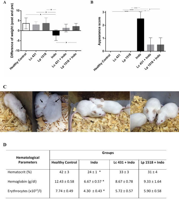
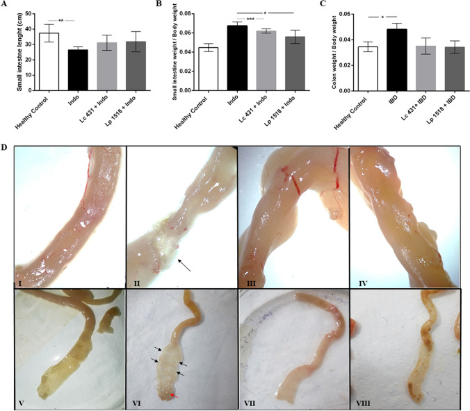
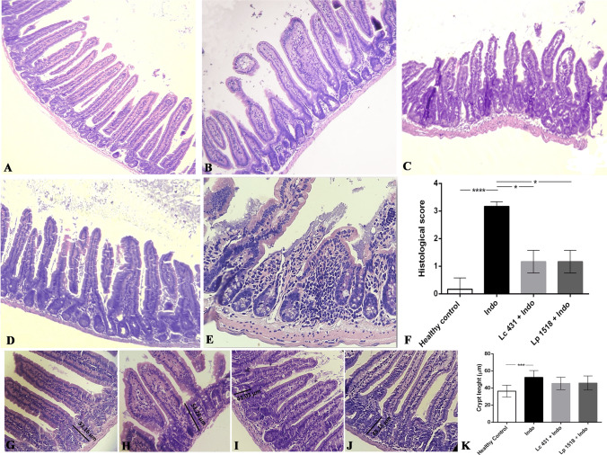
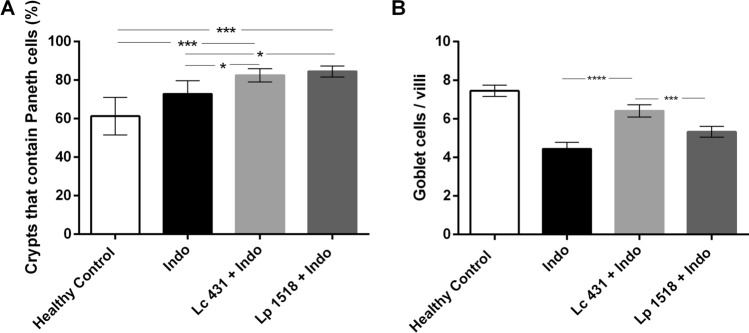
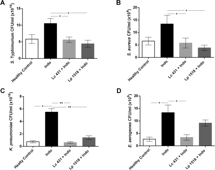
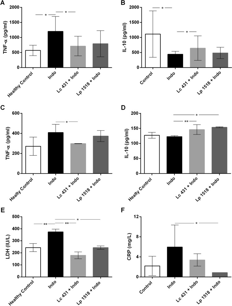
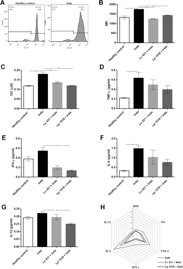
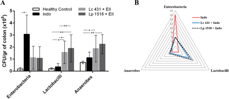
Damage to the small intestine caused by non-steroidal anti-inflammatory drugs (NSAIDs) occurs more frequently than in the upper gastrointestinal tract, is more difficult to diagnose and no effective treatments exist. Hence, we investigated whether probiotics can control the onset of this severe condition in a murine model of intestinal inflammation induced by the NSAID, indomethacin. Probiotic supplementation to mice reduce the body weight loss, anemia, shortening of the small intestine, cell infiltration into the intestinal tissue and the loss of Paneth and Goblet cells associated with intestinal inflammation. Furthermore, a high antimicrobial activity in the intestinal fluids of mice fed with probiotics compared to animals on a conventional diet was elicited against several pathogens. Interestingly, probiotics dampened the oxidative stress and several local and systemic markers of an inflammatory process, as well as increased the secretion of IL-10 by regulatory T cells. Even more importantly, probiotics induced important changes in the large intestine microbiota characterized by an increase in anaerobes and lactobacilli, and a significant decrease in total enterobacteria. We conclude that oral probiotic supplementation in NSAID-induced inflammation increases intestinal antimicrobial activity and reinforces the intestinal epithelial barrier in order to avoid pathogens and commensal invasion and maintain intestinal homeostasis.


Conflict of interest statement

The authors declare no competing interests.

Figures

Figure 1
Figure 1
Clinical features of intestinal inflammation in animals. Balb/c mice supplemented with Lactobacillus casei CRL 431 (Lc 431), Lactobacillus paracasei CNCM I-1518 (Lp 1518), or water, for 7 and 5 days, received indomethacin injections on the last two days. Additional controls fed with Lc 431, Lp 1518 or water, received 2 PBS injections. (A) Body weight differences after and before the indomethacin injection. (B) Appearance score was evaluated according to Shrum et al.. The line at the top of the bars indicates the comparison between the two groups, *p < 0.05. (C) Photography of mice. Panels I- and II- show important patches of piloerected hair pointed by black arrows, in the Indo group. By contrast, the coat is smooth in Lc 431 + Indo and Lp 1518 + Indo groups (Panel III- and IV-, respectively). (D) Evaluation of red blood cells on the day mice were euthanized. Stars show the comparison to the healthy control group. *p < 0.05.
Figure 2
Figure 2
Intestinal damage in mice treated with indomethacin and supplemented with probiotics. Balb/c mice supplemented with Lactobacillus casei CRL 431 (Lc 431), Lactobacillus paracasei CNCM I-1518 (Lp 1518), or water, for 7 and 5 days, received indomethacin on the last two days. On day 8 mice were euthanized and the following parameters were evaluated: (A) small intestine length; (B) the ratio between the weight of the small intestine and the body of each animal; and (C) the ratio between the weight of each large intestine and the body of the animal. The line at the top of the bars indicates the comparison between the two groups. *p < 0.05, **p < 0.01, ***p < 0.001. (D) Representative photography of the small intestine of: Panel I and V- Healthy control; Panel II and VI- Indo; Panel III and VII- Lc 431 + Indo; and Panel IV and VIII- Lp 1518 + Indo mice, revealing the presence of ulcers (black arrows) and a mucosa with granular appearance (red arrows) in mice on a conventional diet treated with indomethacin. I-IV images were taken under observation with a Leica ES2 Stereo Microscope with 10 × High Eyepoint. V-VIII are images of the intestine after cutting them longitudinally.
Figure 3
Figure 3
Hematoxylin and eosin staining of small intestine tissue section. Balb/c mice supplemented with L. casei CRL 431 (Lc 431), L. paracasei CNCM I-1518 (Lp 1518), or water, for 7 and 5 days, received indomethacin injections on the last two days. On day 8 mice were euthanized and samples of the small intestine were taken for histological analysis. Representative micrographs of: (A,G) Healthy control; (B,E,H) mice on a conventional diet treated with indomethacin; (C,I) Lc 431 + Indo; and (D,J) Lp 1518 + Indo. (D) Inflammatory cells infiltrated in indomethacin-treated mice on a conventional diet. (F) Inflammation was qualitatively evaluated according to the number and spread of inflammatory foci from 0 to 4: 0, no leukocytic infiltration; 1, low level of leukocytic infiltration; 2, moderate level of leukocytic infiltration; 3, high level of leukocytic infiltration; and 4, transmural infiltration. *p < 0.05 ***p < 0.001. (K) Length of Lieberkühn crypt in the small intestine of experimental mice. Results (mean ± S.E.M.) are representative of three independent experiments. The line at the top of the bars indicates comparison between the two groups. ***p < 0.01. Magnification: (A-D) 100X, (E, G-J) 400X. Lines in panels G, H, I and J mean: 33.16, 53.26, 43.03 and 48.15 µm, respectively.
Figure 4
Figure 4
Mouse intestinal secretory cells in indomethacin-induced intestinal inflammation. Tissue sections of Balb/c mice orally supplemented with Lactobacillus casei CRL 431 (Lc 431), Lactobacillus paracasei CNCM I-1518 (Lp 1518), or water, for 7 and 5 days and receiving 2 indomethacin injections were stained with hematoxylin and eosin and examined by light microscopy. A blind histological test of the small intestine was performed by analyzing 5 slices of each organ and the percentages of (A) crypts with positive Paneth cells and (B) Goblet cells per villus were determined. Results were expressed as the number of Goblet cells per intestinal villus. The column bar shows a semi-quantitative evaluation for each group. Each bar represents the group mean ± S.E.M. Results are representative of three independent experiments. The line at the top of the bars indicates comparison between the two groups. *p < 0.05, **p < 0.01, ***p < 0.001, ****p < 0.0001.
Figure 5
Figure 5
Antimicrobial activity of intestinal fluids of indomethacin-treated mice. S. aureus (A), S. Typhimirium (B), K. pneumoniae (C) and E. aerogenes (D), (109 CFU/ml), were incubated at 37 °C for 2 h in the presence of the intestinal fluids of: healthy control animals, or mice with induced intestinal inflammation receiving a conventional diet or probiotic supplementation (Indo; Lc 431 + Indo and Lp 1518 + Indo groups, respectively). After co-incubation, viable bacteria were determined by plate count agar. A set of serial dilutions were made and samples of each appropriate dilution were spread on top of solidified agar petri plates. Results (mean ± S.E.M) are representative of three independent experiments. Results were expressed as the differences in CFU/ml after and before the incubation of bacteria with the intestinal fluids. The line at the top of the bars indicates comparison between the two groups. *p < 0.05, **p < 0.01.
Figure 6
Figure 6
Cytokine secretion in the intestinal microenvironment of indomethacin-treated mice fed probiotics. TNF-α (A,C) and IL-10 (B,D) were determined in the (A,B) intestinal fluids and (C,B) supernatant of the epithelial cell culture from healthy control mice and animals with an intestine inflammation process receiving a conventional diet (Indo) or oral supplementation with probiotics (Lc 431 + Indo and Lp 1518 + Indo). Samples were assayed in duplicate by Capture ELISA. The results are representative of 3 independent experiments (n = 6). The line at the top of the bars indicates comparison between the 2 groups. *p < 0.05 and ** p < 0.01.
Figure 7
Figure 7
Release of reactive oxygen species and cytokine mediators of intestinal inflammation by macrophages. Balb/c mice supplemented with L. casei CRL 431 (Lc 431), L. paracasei CNCM I-1518 (Lp 1518), or water, for 7 and 5 days, received indomethacin injections on the last two days. On day 8, mice were euthanized and samples of peritoneal macrophages were taken. ROS production as fluorescence units of oxidized dichlorodihydrofluorescein was detected by flow cytometry and (A) representative histograms and (B) mean fluorescence intensity (MFI) in experimental animals were shown. Peritoneal macrophages were incubated at 37 °C 5% CO2 for 16 h and the supernatant collected for the determination of: (C) NO by Griess reagent; (D,E,F,G) proinflammatory cytokines by Capture ELISA. Results are expressed as mean ± SEM and are representative of at least three independent experiments. The line at the top of the bars indicates comparison between the 2 groups. *p < 0.05 and ** p < 0.01. (H) Radar chart plot of inflammatory mediators released by macrophages from the different experimental groups. Results are expressed as the ratio between each group and the healthy control mice.
Figure 8
Figure 8
Microbial population in the large intestine. Balb/c mice with an indomethacin-induced intestinal inflammatory process receiving a conventional diet (Indo) or oral probiotic supplementation (Lc 431 + Indo and Lp 1518 + Indo) for 7 and 5 days, respectively. At the end of this time, samples of the large intestine were collected and total anaerobic bacteria, lactobacilli, and enterobacterial population were analyzed by plate count agar. (A) Results were expressed as CFU/ml per gram of large intestine (mean ± S.E.M). Three independent experiments were performed. The line at the top of the bars indicates comparison between the two groups. *p < 0.05, **p < 0.01. (B) Mean scores of total bacterial population data in the experimental animals. Results are expressed as the ratio/relationship between each group and healthy controls.

References

    1. Chassaing B, Kumar M, Baker MT, Singh V, Vijay-Kumar M. Mammalian gut immunity. Biomed. J. 2014;37:246–258. doi: 10.4103/2319-4170.130922. - DOI - PMC - PubMed
    1. Artis D. Epithelial-cell recognition of commensal bacteria and maintenance of immune homeostasis in the gut. Nat. Rev. Immunol. 2008;8:411–420. doi: 10.1038/nri2316.Review. - DOI - PubMed
    1. Zasloff M. Antimicrobial peptides of multicellular organisms: my perspective. Adv. Exp. Med. Biol. 2019;1117:3–6. doi: 10.1007/978-981-13-3588-4_1. - DOI - PubMed
    1. Vaishnava S, Behrendt CL, Ismail AS, Eckmann L, Hooper LV. Paneth cells directly sense gut commensals and maintain homeostasis at the intestinal host-microbial interface. Proc. Natl. Acad. Sci. USA. 2008;30:20858–20863. doi: 10.1073/pnas.0808723105. - DOI - PMC - PubMed
    1. Fahlgren A, Hammarstrom S, Danielsson A, Hammarstrom ML. beta-Defensin-3 and -4 in intestinal epithelial cells display increased mRNA expression in ulcerative colitis. Clin. Exp. Immunol. 2004;137:379–385. doi: 10.1111/j.1365-2249.2004.02543.x. - DOI - PMC - PubMed

Publication types

MeSH terms