

Incomplete hippocampal inversion in schizophrenia: prevalence, severity, and impact on hippocampal structure
- PMID: 33437006
- PMCID: PMC8589684
- DOI: 10.1038/s41380-020-01010-z
Incomplete hippocampal inversion in schizophrenia: prevalence, severity, and impact on hippocampal structure
Abstract
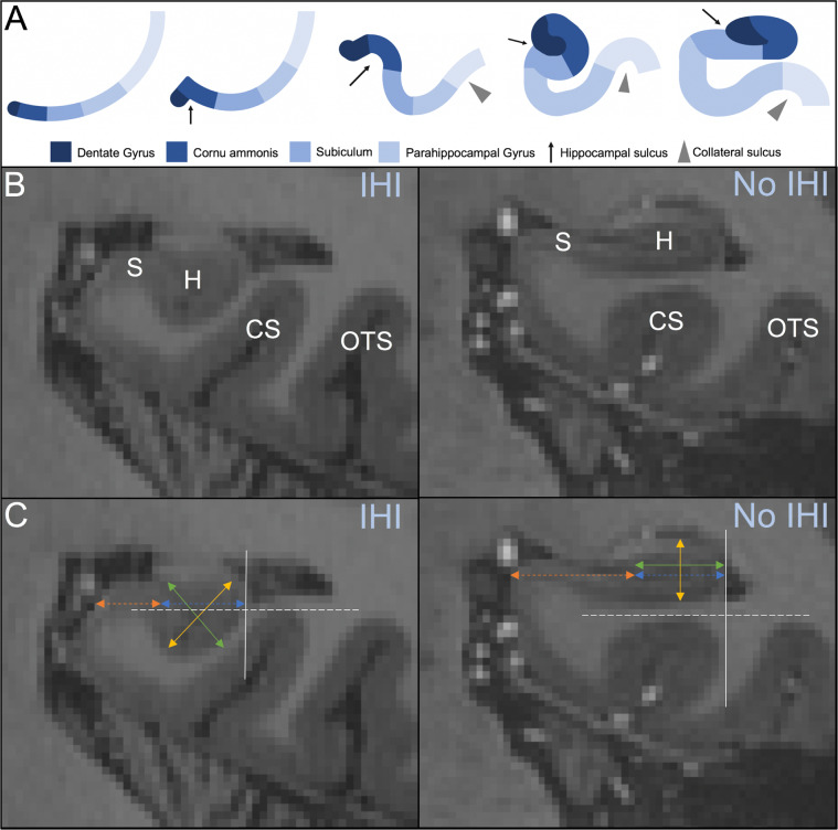
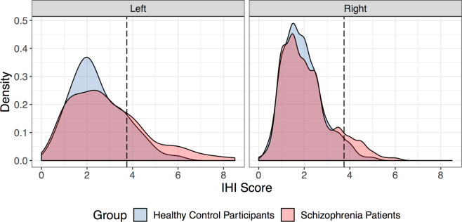
Incomplete hippocampal inversion (IHI) is an anatomical variant of the human brain resulting from an arrest in brain development, especially prevalent in the left hemisphere. We hypothesized that IHI is more common in schizophrenia and contributes to the well-known hippocampal structural differences. We studied 199 schizophrenia patients and 161 healthy control participants with 3 T MRI to establish IHI prevalence and the relationship of IHI with hippocampal volume and asymmetry. IHI was more prevalent (left hemisphere: 15% of healthy control participants, 27% of schizophrenia patients; right hemisphere: 4% of healthy control participants, 10% of schizophrenia patients) and more severe in schizophrenia patients compared to healthy control participants. Severe IHI cases were associated with a higher rate of automated segmentation failure. IHI contributed to smaller hippocampal volume and increased R > L volume asymmetry in schizophrenia. The increased prevalence and severity of IHI supports the neurodevelopmental model of schizophrenia. The impact of this developmental variant deserves further exploration in studies of the hippocampus in schizophrenia.
© 2021. The Author(s), under exclusive licence to Springer Nature Limited part of Springer Nature.
Conflict of interest statement
The authors declare that they have no conflict of interest.
Figures
Similar articles
-
Incomplete Hippocampal Inversion: A Neurodevelopmental Mechanism for Hippocampal Shape Deformation in Schizophrenia.Biol Psychiatry. 2022 Aug 15;92(4):314-322. doi: 10.1016/j.biopsych.2022.02.954. Epub 2022 Feb 23. Biol Psychiatry. 2022. PMID: 35487783 Free PMC article.
-
Hippocampal volume predicts antidepressant efficacy in depressed patients without incomplete hippocampal inversion.Neuroimage Clin. 2016 Apr 27;12:949-955. doi: 10.1016/j.nicl.2016.04.009. eCollection 2016. Neuroimage Clin. 2016. PMID: 27995060 Free PMC article.
-
Incomplete Hippocampal Inversion and Its Relationship to Hippocampal Subfield Volumes and Aging.J Neuroimaging. 2018 Jul;28(4):422-428. doi: 10.1111/jon.12509. Epub 2018 Mar 25. J Neuroimaging. 2018. PMID: 29575376
-
Incomplete hippocampal inversion and epilepsy: A systematic review and meta-analysis.Epilepsia. 2021 Feb;62(2):383-396. doi: 10.1111/epi.16787. Epub 2020 Dec 16. Epilepsia. 2021. PMID: 33325054
-
Hippocampal volume reduction in first-episode and chronic schizophrenia: a review and meta-analysis.Neuroscientist. 2012 Apr;18(2):180-200. doi: 10.1177/1073858410395147. Epub 2011 Apr 29. Neuroscientist. 2012. PMID: 21531988 Review.
Cited by
-
Hippocampal rotation is associated with ventricular atrial size.Pediatr Radiol. 2023 Aug;53(9):1941-1950. doi: 10.1007/s00247-023-05687-6. Epub 2023 May 15. Pediatr Radiol. 2023. PMID: 37183230
-
Automated, open-source segmentation of the Hippocampus and amygdala with the open Vanderbilt archive of the temporal lobe.Magn Reson Imaging. 2021 Sep;81:17-23. doi: 10.1016/j.mri.2021.04.011. Epub 2021 Apr 24. Magn Reson Imaging. 2021. PMID: 33901584 Free PMC article.
-
Automatic rating of incomplete hippocampal inversions evaluated across multiple cohorts.ArXiv [Preprint]. 2025 Jan 20:arXiv:2408.02496v2. ArXiv. 2025. PMID: 39148932 Free PMC article. Updated. Preprint.
-
Epigenome Defines Aberrant Brain Laterality in Major Mental Illnesses.Brain Sci. 2024 Mar 7;14(3):261. doi: 10.3390/brainsci14030261. Brain Sci. 2024. PMID: 38539649 Free PMC article. Review.
-
Lack of correlation between hippocampal substructure atrophy and attention dysfunction in deficit schizophrenia.Schizophrenia (Heidelb). 2023 Apr 20;9(1):24. doi: 10.1038/s41537-023-00354-z. Schizophrenia (Heidelb). 2023. PMID: 37080983 Free PMC article.
References
-
- Haukvik UK, Tamnes CK, Söderman E, Agartz I. Neuroimaging hippocampal subfields in schizophrenia and bipolar disorder: a systematic review and meta-analysis. J Psychiatr Res. 2018;104:217–26. - PubMed
Publication types
MeSH terms
Grants and funding
LinkOut - more resources
Full Text Sources
Other Literature Sources
Medical
