. 2021 Jan 12;4(1):62.
doi: 10.1038/s42003-020-01585-x.

Pharmacological inactivation of the prion protein by targeting a folding intermediate

Affiliations

Pharmacological inactivation of the prion protein by targeting a folding intermediate

Giovanni Spagnolli et al. Commun Biol. .

Abstract

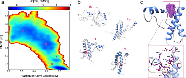
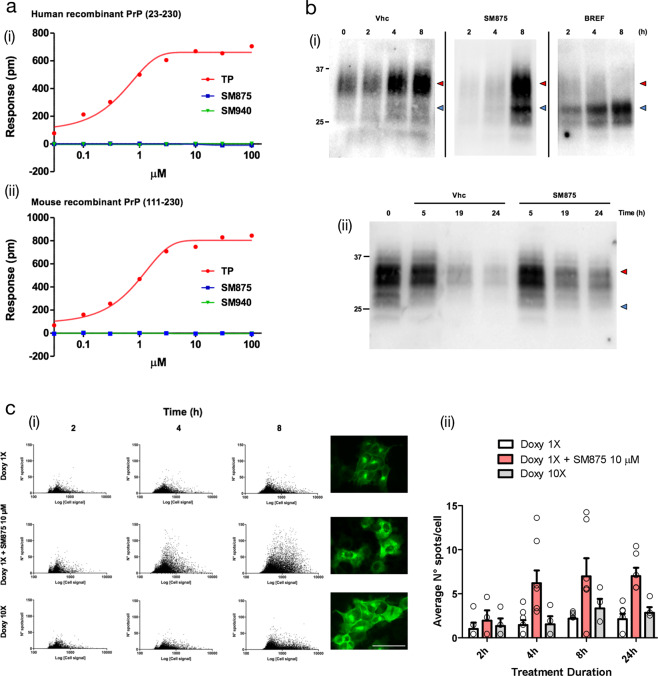
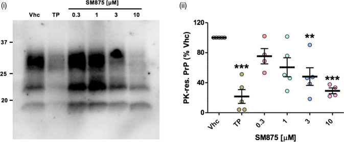
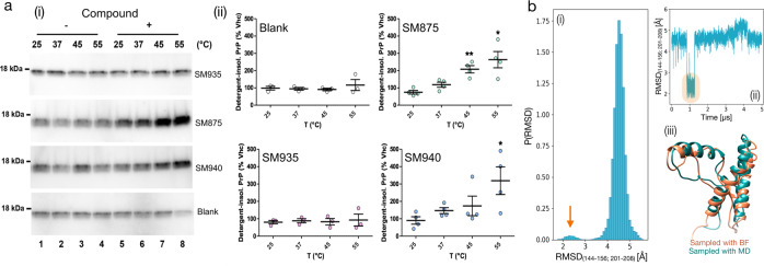
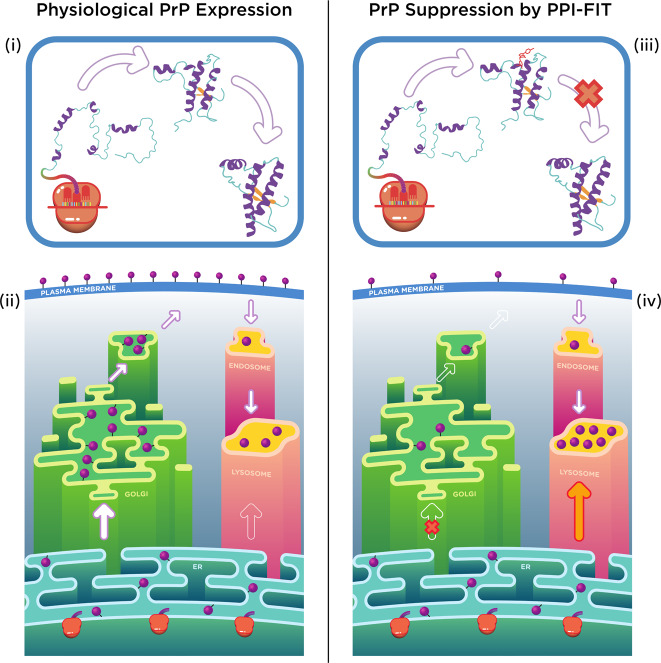
Recent computational advancements in the simulation of biochemical processes allow investigating the mechanisms involved in protein regulation with realistic physics-based models, at an atomistic level of resolution. These techniques allowed us to design a drug discovery approach, named Pharmacological Protein Inactivation by Folding Intermediate Targeting (PPI-FIT), based on the rationale of negatively regulating protein levels by targeting folding intermediates. Here, PPI-FIT was tested for the first time on the cellular prion protein (PrP), a cell surface glycoprotein playing a key role in fatal and transmissible neurodegenerative pathologies known as prion diseases. We predicted the all-atom structure of an intermediate appearing along the folding pathway of PrP and identified four different small molecule ligands for this conformer, all capable of selectively lowering the load of the protein by promoting its degradation. Our data support the notion that the level of target proteins could be modulated by acting on their folding pathways, implying a previously unappreciated role for folding intermediates in the biological regulation of protein expression.


Conflict of interest statement

The authors declare the following competing interests: G.S., G.L., L.M.B., P.F., and E.B. are co-founders and shareholders of Sibylla Biotech SRL. The company exploits the PPI-FIT technology for drug discovery in a wide variety of human pathologies, with the exception of prion diseases.

Figures

Fig. 1
Fig. 1. All-atom reconstruction of the PrP folding pathway.
a Lower-bound approximation of the free energy landscape of the PrP folding obtained from 180 rMD trajectories, plotted as a negative logarithm of the probability distribution expressed as a function of the collective variables Q (fraction of native contacts) and RMSD. The dashed lines delimit the metastable regions of interest (G ≤ 3.7 kBT). b Representative PrP structures of unfolded (U1 and U2), folding intermediate (I) and native conformations (N). Each conformation represents the cluster center of one of the corresponding populated regions (structures depicted in transparency are sampled from the same region, showing conformations variability): the first two (corresponding to the unfolded states U1 and U2) are characterized by RMSD >1.8 nm and a Q < 0.5 (U1), and 1.2 < RMSD < 1.8 nm and a 0.5 < Q < 0.75 (U2); a third one (corresponding to the intermediate folding state I) with 0.55 < RMSD < 0.90 nm and a 0.65 < Q < 0.85; a fourth one (corresponding to the native state N) with RMSD < 0.40 nm and Q > 0.80. c Ribbon diagram of the PrP intermediate highlighting the ligand-binding pocket (purple dots) as identified by SiteMap and DogSiteScorer tools. The purple volume maps the unique druggable pocket identified in the PrP-folding intermediate I. The box shows specific residues defining the site.
Fig. 2
Fig. 2. In vitro validation of selected hits.
HEK293 cells expressing mouse PrP or NEGR-1 were exposed to different concentrations of SM875 a, SM930 b, SM940 c, or SM950 d or vehicle (0.1% DMSO, volume equivalent) for 48 h, lysed in detergent buffer (Tris 10 mM, pH 7.4, 0.5% NP-40, 0.5% TX-100, 150 mM NaCl plus EDTA-free Protease Inhibitors), diluted in Laemmli sample buffer and analyzed by western blotting using anti-PrP (D18) or anti-NEGR-1 (R&D, USA) antibodies. Red arrowheads indicate the expected sizes of mature, fully glycosylated forms of PrP and NEGR-1. The compounds induce a dose-dependent suppression of PrP (i) but not control protein NEGR-1 (ii). The graphs (iii) show the densitometric quantification of the levels of full-length PrP or NEGR-1 from different biologically independent replicates. Each signal was normalized on the corresponding total protein lane (detected by UV of stain-free gels) and expressed as the percentage of the level in vehicle (Vhc)-treated controls (*p < 0.05, **p < 0.01, ***p < 0.005, by one-way ANOVA test). e The picture illustrates the predicted ligand-binding pose of the R (upper panel) and S (lower panel) SM875 enantiomers into the PrP intermediate druggable pocket.
Fig. 3
Fig. 3. SM875 lowers the amount of PrP at a post-translational level in different cell lines.
Cells were exposed to different concentrations of SM875 or vehicle (0.1% DMSO) for 48 h, lysed, and analyzed by western blotting. a In ZR-75 cells, SM875 suppresses PrP, but not Thy-1, in a concentration-dependent fashion. b Similar effects were observed in cultured L929 fibroblasts. c In N2a cells, SM875 shows a dose-dependent lowering effect of PrP at 1–10 μM. However, in contrast to the other cell lines, the compound showed no effect at 30 μM. All signals were detected by using a specific anti-PrP (D18) or anti-Thy-1 primary antibodies. Red arrowheads indicate the expected sizes of mature, fully glycosylated forms of PrP or Thy-1. Western blotting analysis (i) and graphs reporting the densitometric quantification of signals (ii) are shown. Each signal was normalized on the corresponding total protein lane (detected by UV of stain-free gels) and expressed as the percentage of vehicle (Vhc)-treated controls (*p < 0.05, **p < 0.01, ***p < 0.005, by one-way ANOVA test). d Graphs show the levels of PrP mRNA upon treatment with SM875, as evaluated by RT-PCR. Specific forward and reverse primers were used to amplify endogenous or exogenous, mouse or human PrP transcripts (see Materials and Methods). Relative quantification was normalized to mouse or human HPRT (hypoxanthine–guanine phosphoribosyltransferase). Statistical analyses refer to the comparison with vehicle controls (**p < 0.01, ***p < 0.005, by one-way ANOVA test). Dots represent biologically independent replicates. e HEK293 stably expressing a PrP form tagged with a monomerized EGFP molecule at its N-terminus (EGFP-PrP) were incubated with vehicle (0.1% DMSO) control (i) or SM875 at different concentrations (ii–vi) for 24 h. Fluorescence of the EGFP protein was then visualized with an Olympus BX51WI microscope equipped with reflected fluorescence. Scale bar 50 μm.
Fig. 4
Fig. 4. SM875 decreases PrP level via the autophagy-mediated lysosomal degradation.
a (i) PrP-transfected or untransfected HEK293 cells were treated with different concentrations of SM875 or vehicle (0.1% DMSO, volume equivalent) and the levels of PrP, LC3-I, and LC3-II were evaluated by western blotting (indicated by the red arrowheads). As a positive control, cells were treated with 100 μM trehalose (TRE), a disaccharide previously reported to induce autophagy. (ii) Graphs show the densitometric quantification of LC3-II from independent replicates (n = 8 for PrP-expressing HEK293 cells; n = 5 for untransfected HEK293 cells). Each signal was normalized on the corresponding total protein lane (detected by UV) and expressed as the percentage of vehicle (Vhc)-treated controls (**p < 0.01, by one-way ANOVA test). b (i) ZR-75 cells were treated with different concentrations of SM875 in the presence or absence of autophagy–lysosomal inhibitor bafilomycin A1 (BAF, 10 μM) and PrP levels were evaluated by western blotting. (ii) Graphs show the densitometric quantification of full-length PrP from independent replicates (n = 3). Each signal was normalized on the corresponding total protein lane (detected by UV) and expressed as the percentage of vehicle (Vhc)-treated controls (**p < 0.01, by one-way ANOVA test).
Fig. 5
Fig. 5. SM875 acts exclusively on non-native, newly synthesized PrP.
a DMR was employed to assess whether SM875 has an affinity for the native conformation of PrP. Different concentrations (0.03–100 μM) of SM875, SM940 or PrP ligand Fe3+-TMPyP (TP), used as a control, were added to label-free microplate well surfaces on which either (i) full-length, human recombinant PrP (23–230) or (ii) N-terminally deleted mouse recombinant PrP (111–230) had previously been immobilized. All signals were fitted (continuous lines), when possible, to a sigmoidal function using a 4PL non-linear regression model. In contrast to SM875 and SM940, TP shows a detectable affinity for both full-length, human, and N-terminally deleted, mouse PrP molecules (for full-length PrP, Kd = 0.67 ± 0.05, R2 = 0.99). b In order to dissect the effect of SM875 on nascent vs mature, native PrP molecules, we turned to RK13 cells expressing mouse PrP under control a doxycycline-inducible promoter. (i) PrP expression was induced over 8 h, in the presence of SM875 (10 μM), brefeldin-1A (BREF, 10 μM), or vehicle (Vhc) control, samples were collected at different time points (indicated) and PrP signals were visualized by western blotting. Signals were detected by probing membrane blots with anti-PrP antibody (D18). As expected, in control cells, the level of full-length PrP (red arrowheads) increases in a time-dependent fashion. Conversely, a lower molecular weight band (blue arrowheads) is detected in brefeldin-treated cells. (ii) Next, we designed an experiment to test the effect of SM875 exclusively on mature, natively folded PrP. PrP expression was induced for 24 h, in the absence of any additional treatment. Doxycycline was then removed, and after 4 h without inducer, the cells were exposed to SM875 or Vhc control, and subsequently lysed at different time points (indicated). In this experimental setting, cells are exposed to SM875 only when all PrP molecules are synthesized and likely in transit to, or already reached the plasma membrane. In these conditions, normal PrP patterns appear in both compound-treated and Vhc-treated cells. c A high-content approach was employed to the same experimental setting described above by analyzing the localization of PrP after immunostaining with an anti-PrP antibody (D18) coupled to an Alexa 488 secondary antibody. (i) The expression of PrP was induced for 2, 4, or 8 h by doxycycline 1× (0.01 mg/mL) or 10× (0.1 mg/mL) and the effect of SM875 (10 μM) incubated with doxycycline 1× was measured by Harmony software after the image acquisition performed by Operetta Imaging System. Green spots detected in cells were quantified and plotted against the total green fluorescence relative to each cell. Representative images were acquired at 8 h of incubation, scale bar 50 μm. (ii) Quantification of the average number of green spots per cell in wells incubated for 2, 4, 8, and 24 h with doxycycline 1× (white bars), doxycycline 10× (gray bars) and doxycycline 1× + SM875 (red bars). Quantification of at least three independent experiments.
Fig. 6
Fig. 6. SM875 inhibits prion replication in L929 mouse fibroblasts.
L929 mouse fibroblasts were infected with the RML prion strain and then propagated for five sequential passages before exposure to SM875 (indicated concentrations), anti-prion compound Fe3+-TMPyP (TP, 10 μM) or vehicle (0.1% DMSO) for 48 h. PrPSc loads were then estimated by treating cell lysates with PK and analyzing PrP content by western blotting (i). Signals were detected by probing membrane blots with anti-PrP antibody (D18). Results show that SM875 inhibits prion replication in a dose-dependent fashion, with the maximal effect (obtained at 10 μM) comparable to that of TP; (ii) the graph shows the densitometric quantification of all PK-resistant PrP bands from independent replicates (n ≥ 4). Quantification was obtained by densitometric analysis of the western blots, normalizing each signal on the corresponding PK-untreated lane and expressed as the percentage of vehicle (Vhc)-treated controls (**p < 0.01, *** p < 0.005, by one-way ANOVA test).
Fig. 7
Fig. 7. SM875 induces the aggregation of PrP in a temperature-dependent fashion.
a Recombinant PrP (111–231) was diluted in a detergent buffer (final concentration of 0.5 μM), placed at one of the indicated temperatures and incubated for 1 h with either vehicle (Vhc, lanes 1–4) or (lanes 5–8) assay buffer (blank), SM875, SM940, or SM935 (10 μM). After incubation, samples were subjected to ultracentrifugation, and the resulting detergent-insoluble pellets, corresponding to the aggregated fraction of PrP, were analyzed by western blotting (i). Signals were detected by probing membrane blots with anti-PrP antibody (D18). (ii) Graphs show the densitometric quantification of recombinant PrP bands from independent replicates. Each signal was normalized and expressed as the percentage of the corresponding Vhc-treated sample (*p < 0.05, **p < 0.01, by one-way ANOVA test). b Native PrP (residues 125–228, PDB 1QLX) was subjected to 21 μs of cumulative (3 × 5 μs + 2 × 3 μs) MD simulations at 310 K. (i) The RMSD of residues lying within the contact region between helix-1 and helix-3 (indicated) was computed with respect to the structure of the previously identified PrP-folding intermediate. (i) The graph shows the probability distribution of the RMSD. The orange arrow indicates the population of PrP conformations resembling the folding intermediate. (ii) Individual trajectory exploring the PrP-folding intermediate from the native state (highlighted in orange). (iii) Superimposition of the PrP-folding intermediate identified by the BF method and the conformation reached from the native state, as observed by plain MD.
Fig. 8
Fig. 8. Model for the PPI-FIT-based suppression of PrP.
The model schematically illustrates the rationale underlying the PPI-FIT method applied to PrP. (i and iii) The schematics highlight PrP folding in the presence or absence of a small molecule targeting the PrP intermediate. (ii) PrP follows a typical expression pathway for GPI-anchored proteins. The polypeptide is directly synthesized into the lumen of the ER, once properly folded and post-translationally modified it traffics to the Golgi apparatus, where sugar moieties are matured, and then delivered to the cell surface. From the plasma membrane, PrP molecules could enter into the endosomal recycling pathways, and eventually recycled into the lysosomes. (iv) Aberrantly folded conformers of PrP, as the case for some previously reported mutant, could be re-routed directly from ER to the lysosomes. In the PPI-FIT method, a small molecule interferes with PrP expression by binding to a folding intermediate, which gets delivered to the lysosomes, leading to an overall decrease of the protein at the cell surface.

References

    1. Balch WE, Morimoto RI, Dillin A, Kelly JW. Adapting proteostasis for disease intervention. Science. 2008;319:916–919. doi: 10.1126/science.1141448. - DOI - PubMed
    1. Anfinsen CB, Scheraga HA. Experimental and theoretical aspects of protein folding. Adv. Protein Chem. 1975;29:205–300. doi: 10.1016/S0065-3233(08)60413-1. - DOI - PubMed
    1. Steinmetz AC, Renaud JP, Moras D. Binding of ligands and activation of transcription by nuclear receptors. Annu Rev. Biophys. Biomol. Struct. 2001;30:329–359. doi: 10.1146/annurev.biophys.30.1.329. - DOI - PubMed
    1. Powers ET, Morimoto RI, Dillin A, Kelly JW, Balch WE. Biological and chemical approaches to diseases of proteostasis deficiency. Annu Rev. Biochem. 2009;78:959–991. doi: 10.1146/annurev.biochem.052308.114844. - DOI - PubMed
    1. Chiti F, Dobson CM. Protein misfolding, functional amyloid, and human disease. Annu Rev. Biochem. 2006;75:333–366. doi: 10.1146/annurev.biochem.75.101304.123901. - DOI - PubMed

Publication types

MeSH terms