

Minocycline Alleviates Cluster Formation of Activated Microglia and Age-dependent Dopaminergic Cell Death in the Substantia Nigra of Zitter Mutant Rat
- PMID: 33437100
- PMCID: PMC7785462
- DOI: 10.1267/ahc.20-00022
Minocycline Alleviates Cluster Formation of Activated Microglia and Age-dependent Dopaminergic Cell Death in the Substantia Nigra of Zitter Mutant Rat
Abstract
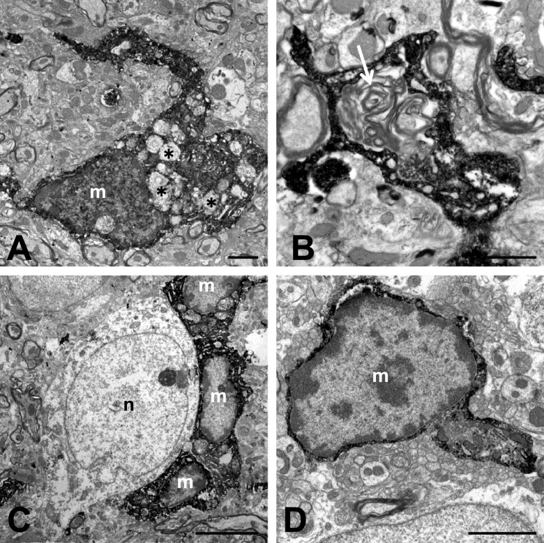
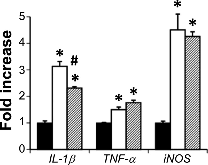
Microglial activation is a component of neurodegenerative pathology. Here, we examine whether activated microglia participate in age-related dopaminergic (DA) cell death in the substantia nigra pars compacta (SNc) of the zitter (zi/zi) rat, a mutant characterized by deletion of the attractin gene. Confocal microscopy with double-immunohistochemical staining revealed activated microglia-formed cell-clusters surrounding DA neurons in the SNc from 2 weeks after birth. An immunoelectron microscopic study showed that the cytoplasm of activated microglia usually contains phagosome-like vacuoles and lamellar inclusions. Expression levels of the pro-inflammatory cytokines interleukin-1β (IL-1β), tumor necrosis factor-α (TNF-α) and inducible nitric oxide synthase (iNOS) were increased in the midbrain of 2-month-old zi/zi rats. Chronic treatment with the anti-inflammatory agent minocycline altered the morphology of the microglia, reduced cluster formation by the microglia, and attenuated DA cell death in the SNc, and reduced the expression of IL-1β in the midbrain. These results indicate that activated microglia, at least in part and especially at the initial phase, contribute to DA cell death in the SNc of the zi/zi rat.
Keywords: activated microglia; dopamine neuron; minocycline; pro-inflammatory cytokines; substantia nigra.
2020 The Japan Society of Histochemistry and Cytochemistry.
Conflict of interest statement
VThe authors declare that they have no conflicts of interest.
Figures


Similar articles
-
Role of neuronal nitric oxide synthase in slowly progressive dopaminergic neurodegeneration in the Zitter rat.Nitric Oxide. 2018 Aug 1;78:41-50. doi: 10.1016/j.niox.2018.05.007. Epub 2018 May 22. Nitric Oxide. 2018. PMID: 29792933
-
Neuroprotective effects of melatonin on the nigrostriatal dopamine system in the zitter rat.Neurosci Lett. 2012 Jan 6;506(1):79-83. doi: 10.1016/j.neulet.2011.10.053. Epub 2011 Oct 28. Neurosci Lett. 2012. PMID: 22056485
-
Degeneration of dopaminergic neurons in the substantia nigra of zitter mutant rat and protection by chronic intake of Vitamin E.Neurosci Lett. 2005 Jun 3;380(3):252-6. doi: 10.1016/j.neulet.2005.01.053. Epub 2005 Feb 8. Neurosci Lett. 2005. PMID: 15862896
-
Manganese induces dopaminergic neurodegeneration via microglial activation in a rat model of manganism.Toxicol Sci. 2009 Jan;107(1):156-64. doi: 10.1093/toxsci/kfn213. Epub 2008 Oct 4. Toxicol Sci. 2009. PMID: 18836210
-
Inhibition of prothrombin kringle-2-induced inflammation by minocycline protects dopaminergic neurons in the substantia nigra in vivo.Neuroreport. 2014 May 7;25(7):489-95. doi: 10.1097/WNR.0000000000000122. Neuroreport. 2014. PMID: 24488033
Cited by
-
Effects of Smart Drugs on Cholinergic System and Non-Neuronal Acetylcholine in the Mouse Hippocampus: Histopathological Approach.J Clin Med. 2022 Jun 9;11(12):3310. doi: 10.3390/jcm11123310. J Clin Med. 2022. PMID: 35743382 Free PMC article.
-
Emerging role of microglia in the developing dopaminergic system: Perturbation by early life stress.Neural Regen Res. 2026 Jan 1;21(1):126-140. doi: 10.4103/NRR.NRR-D-24-00742. Epub 2024 Nov 13. Neural Regen Res. 2026. PMID: 39589170 Free PMC article.
References
-
- Brown G. C. and Bal-Price A. (2003) Inflammatory neurodegeneration mediated by nitric oxide, glutamate, and mitochondria. Mol. Neurobiol. 27; 325–355. - PubMed
-
- Cicchetti F., Brownell A. L., Williams K., Chen Y. I., Livni E. and Isacson O. (2002) Neuroinflammation of the nigrostriatal pathway during progressive 6-OHDA dopamine degeneration in rats monitored by immunohistochemistry and PET imaging. Eur. J. Neurosci. 15; 991–998. - PubMed
-
- Dong Y. and Benveniste E. N. (2001) Immune function of astrocytes. Glia 36; 180–190. - PubMed
LinkOut - more resources
Full Text Sources
Miscellaneous