

Salinomycin suppresses TGF-β1-induced EMT by down-regulating MMP-2 and MMP-9 via the AMPK/SIRT1 pathway in non-small cell lung cancer
- PMID: 33437206
- PMCID: PMC7797542
- DOI: 10.7150/ijms.50080
Salinomycin suppresses TGF-β1-induced EMT by down-regulating MMP-2 and MMP-9 via the AMPK/SIRT1 pathway in non-small cell lung cancer
Abstract
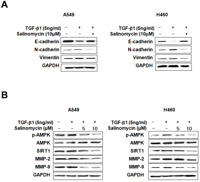
Salinomycin (Sal) is a recently identified anti-tumor drug for treating several types of solid tumor; however, its effects on the migratory and invasive properties of non-small cell lung cancer (NSCLC) remain unclear. This study investigated the inhibitory effect underlying mechanisms of Salon transforming growth factor-β1 (TGF-β1)-induced epithelial-to-mesenchymal transition (EMT) and cell migration. Sal solidly blocked cell migration and invasion enhancement by TGF-β1-induced EMT, through recovering E-cadherin loss and suppressing mesenchymal markers induction, as well as TGF-β1-mediated AMPK/SIRT signaling activity upregulation. The pharmacologic inhibition or knockdown of AMPK or SIRT1 can act synergistically with Sal to inhibit TGF-β1-induced MMP-2 and MMP-9. In contrast, AMPK or SIRT1 upregulation can protect against TGF-β1-induced MMP-2 and MMP-9 inhibition by Sal. Next we demonstrated that the MMP-2 and MMP-9 knockdown can act synergistically with Sal to inhibit TGF-β1-induced EMT. Moreover, treatment of PMA of MMP activator increased TGF-β1-induced MMP-2 and MMP-9, even with Sal. Our results demonstrate that Sal suppresses TGF-β1-induced EMT by downregulating MMP-2 and MMP-9 through the AMPK/SIRT pathway, thereby inhibiting lung cancer cell migration and invasion.
Keywords: AMPK; EMT; Lung cancer; MMP; SIRT; Salinomycin; TGF-β1.
© The author(s).
Conflict of interest statement
Competing Interests: The authors have declared that no competing interest exists.
Figures


Similar articles
-
Celecoxib and sulindac inhibit TGF-β1-induced epithelial-mesenchymal transition and suppress lung cancer migration and invasion via downregulation of sirtuin 1.Oncotarget. 2016 Aug 30;7(35):57213-57227. doi: 10.18632/oncotarget.11127. Oncotarget. 2016. PMID: 27528025 Free PMC article.
-
Kaempferol Suppresses Transforming Growth Factor-β1-Induced Epithelial-to-Mesenchymal Transition and Migration of A549 Lung Cancer Cells by Inhibiting Akt1-Mediated Phosphorylation of Smad3 at Threonine-179.Neoplasia. 2015 Jul;17(7):525-37. doi: 10.1016/j.neo.2015.06.004. Neoplasia. 2015. PMID: 26297431 Free PMC article.
-
Ursolic Acid Attenuates TGF-β1-Induced Epithelial-Mesenchymal Transition in NSCLC by Targeting Integrin αVβ5/MMPs Signaling.Oncol Res. 2019 May 7;27(5):593-600. doi: 10.3727/096504017X15051723858706. Epub 2017 Sep 14. Oncol Res. 2019. PMID: 28911340 Free PMC article.
-
AMPK: An energy sensor for non-small cell lung cancer progression and treatment.Pharmacol Res. 2025 Feb;212:107592. doi: 10.1016/j.phrs.2025.107592. Epub 2025 Jan 11. Pharmacol Res. 2025. PMID: 39805353 Review.
-
Dichloroacetate and Salinomycin as Therapeutic Agents in Cancer.Med Sci (Basel). 2025 Apr 23;13(2):47. doi: 10.3390/medsci13020047. Med Sci (Basel). 2025. PMID: 40407542 Free PMC article. Review.
Cited by
-
Roles of MMP-2 and MMP-9 and their associated molecules in the pathogenesis of keloids: a comprehensive review.Front Pharmacol. 2024 Nov 25;15:1444653. doi: 10.3389/fphar.2024.1444653. eCollection 2024. Front Pharmacol. 2024. PMID: 39654616 Free PMC article. Review.
-
MicroRNA-195-5p suppresses the proliferation, migration, invasion and epithelial-mesenchymal transition of laryngeal cancer cells in vitro by targeting E2F3.Exp Ther Med. 2021 Oct;22(4):1078. doi: 10.3892/etm.2021.10512. Epub 2021 Jul 28. Exp Ther Med. 2021. PMID: 34447471 Free PMC article.
-
Non-small cell lung cancer map and analysis: exploring interconnected oncogenic signal integrators.Mamm Genome. 2025 Jun;36(2):573-600. doi: 10.1007/s00335-025-10110-6. Epub 2025 Feb 12. Mamm Genome. 2025. PMID: 39939487
-
The Impact of Catalpol on Proliferation, Apoptosis, Migration, and Oxidative Stress of Lung Cancer Cells Based on Nrf2/ARE Signaling.Biomed Res Int. 2022 Jul 18;2022:5621341. doi: 10.1155/2022/5621341. eCollection 2022. Biomed Res Int. 2022. Retraction in: Biomed Res Int. 2024 Jan 9;2024:9860506. doi: 10.1155/2024/9860506. PMID: 35898682 Free PMC article. Retracted.
-
Targeting MAPK Signaling: Loureirins A and B from Dracaena Loureiri Inhibit Epithelial-Mesenchymal Transition and Invasion in Non-Small Cell Lung Cancer Cell Lines.Life (Basel). 2025 Mar 3;15(3):396. doi: 10.3390/life15030396. Life (Basel). 2025. PMID: 40141741 Free PMC article.
References
MeSH terms
Substances
LinkOut - more resources
Full Text Sources
Other Literature Sources
Medical
Miscellaneous
