. 2020 Dec 15;12(12):7995-8006.
eCollection 2020.

Decreased expression of adenosine receptor 2B confers cardiac protection against ischemia via restoring autophagic flux

Affiliations

Decreased expression of adenosine receptor 2B confers cardiac protection against ischemia via restoring autophagic flux

Enbo Zhan et al. Am J Transl Res. .

Abstract

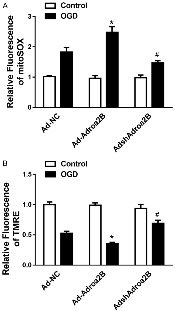
Adora2B (adenosine receptor 2B) has been reported as one of the key modulators during cardiac remodeling after acute myocardial infarction (AMI). However, the molecular mechanism involved has not been well investigated. Thus, our study aims to investigate whether Adora2B contributes to cardiac remodeling after AMI and its underlying mechanisms. Adenovirus harboring Adora2B or shAdora2B was injected in the border zone in a mouse model of AMI experimentally produced by permanent ligation of left anterior descending (LAD) coronary artery. Decreased Adora2B expression protected the cardiomyocytes from MI-induced autophagic flux obstacle, improved cardiac function, and reduced fibrosis after MI. Adora2B downregulation attenuated the accumulation of LC3-II and p62, which are autophagy substrate proteins. An adenovirus containing mRFP-GFP-LC3 showed that decreased expression of Adora2B restored the autophagic flux by enhancing autophagosome conversion to autophagolysosome. Also, Adora2B knockdown improved cardiomyocytes' survival and protected mitochondrial function of cardiomyocytes insulted with hypoxia. Notably, the effect of Adora2B on autophagy flux and cardiomyocyte protection could be mitigated by autophagy inhibitor chloroquine. Our results demonstrate that decreased expression of Adora2B protected cardiomyocytes from impaired autophagy flux induced by MI. Modulation Adora2B expression plays a significant role in blunting the worsening of heart function and reducing scar formation, suggesting therapeutic potential by targeting Adora2B in AMI for the infarct healing.

Keywords: Acute myocardial infarction; Adora2B; apoptosis; autophagy flux.


Conflict of interest statement

None.

Figures

Figure 1
Figure 1
Adora2B was induced in the heart infarct border after LAD ligation and in the neonatal rat cardiomyocytes (NRCMs) after oxygen-glucose deprivation (OGD) injury. A. Representative immunoblotting images and analysis result depicting p62 and Adora2B in the heart infarct border at different time points after LAD ligation. B. Representative western blotting image and analysis depicting p62 and Adora2B in the NRCMs for different time points after OGD treatment. C. Quantitative PCR analysis of the relative abundance of p62 and Adora2B in the heart infarct border at different time points after MI. D. Quantitative PCR analysis of the relative abundance of p62 and Adora2B in the NRCMs for different time points after OGD treatment.
Figure 2
Figure 2
Adora2B critically regulates cardiomyocytes’ survival during OGD. (A) Western blot assay was performed to confirm that the Adora2B expression. (B) The cell viability was analyzed by CCK8 assay with or without OGD treatment on NRCMs transfected with adenovirus carrying Adora2B or shRNA (*P < 0.05 vs. Ad-Null; #P < 0.05 vs. Si-Scramble, n = 5). (C) The representative image and analysis of flow cytometry showing that Adora2B overexpression attenuated cardiomyocytes injury, whereas Adora2B knockdown (D) aggravated cardiomyocytes injury after OGD treatment. *P < 0.05 vs. Ad-Null; #P < 0.05 vs. Si-Scramble, n = 5.
Figure 3
Figure 3
Adora2B critically regulates cardiomyocyte’s mitochondrial function during OGD. Cumulative data of assessing mitochondrial ROS generation (A) and mitochondrial membrane potential (B) in NRCMs suffered from OGD for 12 hours. *P < 0.05 vs. control; #P < 0.05 vs. Ad-scramble-OGD group.
Figure 4
Figure 4
Decreased Adora2B attenuated reverse LV remodeling after MI in mice. A, B. The representative images and analysis results of echocardiographic assessment of hearts 28 days after MI. C. The representative images and analysis results of Masson staining assessment of the hearts 28 days after MI. *P < 0.05 vs. control; #P < 0.05 vs. Ad-NC-CQ group.
Figure 5
Figure 5
Decreased Adora2B confers myocardial protection by restoring autophagy flux in vitro. A. The autophagy associated protein marker p62 and LC3 I/II was examined by Western blotting in the heart infarct border 28 days after MI with or without CQ. B. The representative fluorescent image and analysis of the autophagosome and autophagolysosome in the presence or absence of CQ after Ad-shAdora2B transfection in the NRCMs with OGD treatment. (*P < 0.05 vs. OGD + Ad-Null; #P < 0.05 vs. OGD + Ad-shAdora2B, n = 5).
Figure 6
Figure 6
Decreased Adora2B promotes cardiomyocytes survival against OGD is mediated by restoring autophagy flux. A. The representative image and analysis of flow cytometry showing that cardio-protection of decreased Adora2B expression was ameliorated after CQ administration. B. The representative immunoblotting image and analysis of cleaved-Caspase 3 expressions showing that cardio-protection of decreased Adora2B expression was ameliorated after CQ administration. (*P < 0.05 vs. CTL; #P < 0.05 vs. OGD + Ad-Null; &P < 0.05 vs. OGD + Ad-shAdora2B, n = 6).

Similar articles

Cited by

References

    1. Ruparelia N, Godec J, Lee R, Chai JT, Dall’Armellina E, McAndrew D, Digby JE, Forfar JC, Prendergast BD, Kharbanda RK, Banning AP, Neubauer S, Lygate CA, Channon KM, Haining NW, Choudhury RP. Acute myocardial infarction activates distinct inflammation and proliferation pathways in circulating monocytes, prior to recruitment, and identified through conserved transcriptional responses in mice and humans. Eur Heart J. 2015;36:1923–34. - PMC - PubMed
    1. Frangogiannis NG. Inflammation in cardiac injury, repair and regeneration. Curr Opin Cardiol. 2015;30:240–5. - PMC - PubMed
    1. Nakai A, Yamaguchi O, Takeda T, Higuchi Y, Hikoso S, Taniike M, Omiya S, Mizote I, Matsumura Y, Asahi M, Nishida K, Hori M, Mizushima N, Otsu K. The role of autophagy in cardiomyocytes in the basal state and in response to hemodynamic stress. Nat Med. 2007;13:619. - PubMed
    1. Nishida K, Taneike M, Otsu K. The role of autophagic degradation in the heart. J Mol Cell Cardiol. 2015;78:73–79. - PubMed
    1. Ma X, Liu H, Foyil SR, Godar RJ, Weinheimer CJ, Hill JA, Diwan A. Impaired autophagosome clearance contributes to cardiomyocyte death in ischemia/reperfusion injury. Circulation. 2012;125:3170–3181. - PMC - PubMed

LinkOut - more resources