

Effect of book-shaped acellular tendon scaffold with bone marrow mesenchymal stem cells sheets on bone-tendon interface healing
- PMID: 33437635
- PMCID: PMC7773951
- DOI: 10.1016/j.jot.2020.02.013
Effect of book-shaped acellular tendon scaffold with bone marrow mesenchymal stem cells sheets on bone-tendon interface healing
Abstract
Background: Tissue engineering has exhibited great effect on treatment for bone-tendon interface (BTI) injury. The aim of this study was to evaluate the effect of a book-shaped acellular tendon scaffold (ATS) with bone marrow mesenchymal stem cells sheets (MSCS) for BTI injury repair.
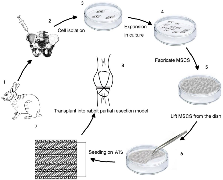
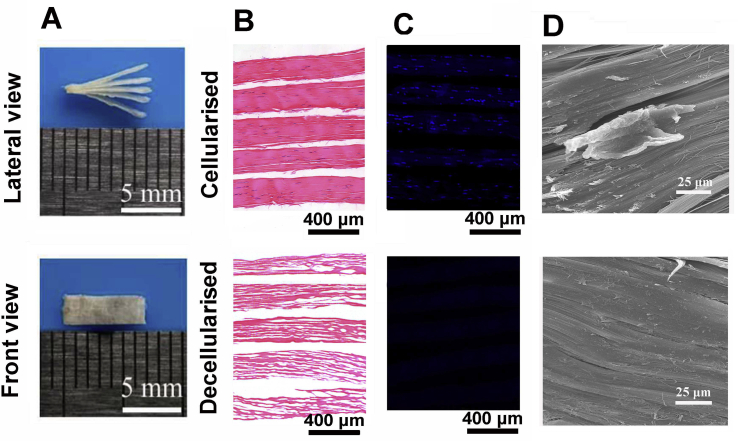
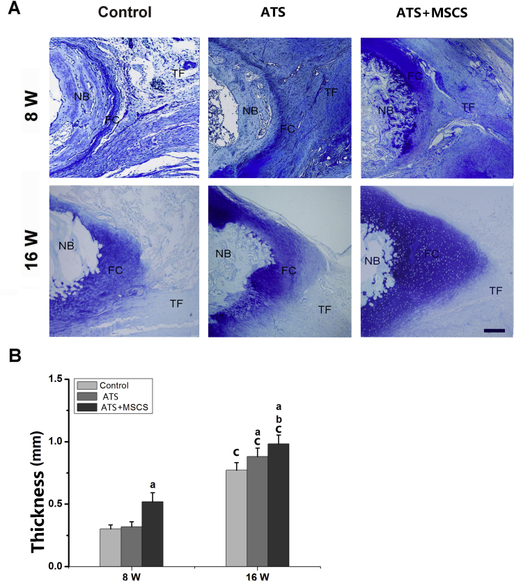
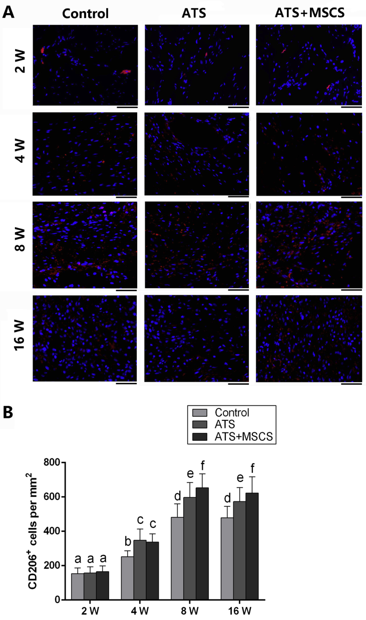
Methods: ATS was designed based on the shape of "book", decellularization effect was evaluated by Hematoxylin and eosin (H&E), 4', 6-diamidino-2-phenylindole (DAPI) and scanning electron microscopy (SEM), then bone marrow mesenchymal stem cells (MSCs) were cultured on ATS to assess the differentiation inductivity of ATS. A rabbit right partial patellotomy model was established, and MSCS seeded on ATS were implanted into the lesion site. The patella-patellar tendon (PPT) at 2, 4, 8 or 16 weeks post-operation were obtained for histological, biomechanical and immunofluorescence analysis.
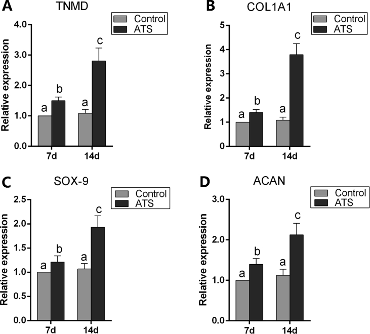
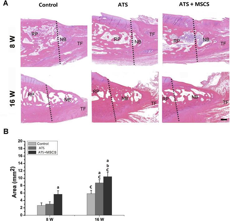
Results: H&E, DAPI and SEM results confirmed the efficiency of decellularization of ATS, and their in vitro tenogenic and chondrogenic ability were successfully identified. In vivo results showed increased macrophage polarization toward the M2 phenotype, IL-10 expression, regenerated bone and fibrocartilage at the patella-patellar tendon interface of animals received MSCS modified ATS implantation. In addition, the level of tensile strength was also the highest in MSCS modified ATS implantation group.
Conclusion: This study suggests that ATS combined with MSCS performed therapeutic effects on promoting the regeneration of cartilage layer and enhancing the healing quality of patella-patellar tendon interface.
The translational potential of this article: This study showed the good biocompatibility of the ATS, as well as the great efficacy of ATS with MSCS on tendon to bone healing. The results meant that the novel book-shaped ATS with MSCS may have a great potential for clinical application.
Keywords: Bone marrow mesenchymal stem cell sheets; Book-shaped acellular tendon scaffold; Patella-patellar tendon interface; Regeneration; Tissue engineering.
© 2020 The Author(s).
Figures
Similar articles
-
-Book-shaped decellularized tendon matrix scaffold combined with bone marrow mesenchymal stem cells-sheets for repair of achilles tendon defect in rabbit.J Orthop Res. 2019 Apr;37(4):887-897. doi: 10.1002/jor.24255. Epub 2019 Mar 28. J Orthop Res. 2019. PMID: 30816590
-
Structure and ingredient-based biomimetic scaffolds combining with autologous bone marrow-derived mesenchymal stem cell sheets for bone-tendon healing.Biomaterials. 2020 May;241:119837. doi: 10.1016/j.biomaterials.2020.119837. Epub 2020 Feb 18. Biomaterials. 2020. PMID: 32109704
-
Comparative Evaluation of the Book-Type Acellular Bone Scaffold and Fibrocartilage Scaffold for Bone-Tendon Healing.J Orthop Res. 2019 Aug;37(8):1709-1722. doi: 10.1002/jor.24301. Epub 2019 Apr 24. J Orthop Res. 2019. PMID: 30977542
-
Mesenchymal stem cells and macrophages and their interactions in tendon-bone healing.J Orthop Translat. 2023 Jan 20;39:63-73. doi: 10.1016/j.jot.2022.12.005. eCollection 2023 Mar. J Orthop Translat. 2023. PMID: 37188000 Free PMC article. Review.
-
Tendon-Bone Healing: Synergistic Role of Platelets and Mesenchymal Stem Cells in Tissue Engineering.Tissue Eng Part B Rev. 2025 May 22. doi: 10.1089/ten.teb.2024.0333. Online ahead of print. Tissue Eng Part B Rev. 2025. PMID: 40402854 Review.
Cited by
-
Structure, ingredient, and function-based biomimetic scaffolds for accelerated healing of tendon-bone interface.J Orthop Translat. 2024 Jul 31;48:70-88. doi: 10.1016/j.jot.2024.07.007. eCollection 2024 Sep. J Orthop Translat. 2024. PMID: 39185339 Free PMC article.
-
Combined Verapamil-Polydopamine Nanoformulation Inhibits Adhesion Formation in Achilles Tendon Injury Using Rat Model.Int J Nanomedicine. 2023 Jan 6;18:115-126. doi: 10.2147/IJN.S377600. eCollection 2023. Int J Nanomedicine. 2023. PMID: 36636643 Free PMC article.
-
Effects of Platelet-Rich Plasma on Tendon-Bone Healing After Anterior Cruciate Ligament Reconstruction.Orthop Surg. 2022 Jan;14(1):88-95. doi: 10.1111/os.13175. Epub 2021 Dec 6. Orthop Surg. 2022. PMID: 34870370 Free PMC article.
-
Hierarchy Reproduction: Multiphasic Strategies for Tendon/Ligament-Bone Junction Repair.Biomater Res. 2025 Jan 22;29:0132. doi: 10.34133/bmr.0132. eCollection 2025. Biomater Res. 2025. PMID: 39844867 Free PMC article. Review.
-
Engineering an enthesis-like graft for rotator cuff repair: An approach to fabricate highly biomimetic scaffold capable of zone-specifically releasing stem cell differentiation inducers.Bioact Mater. 2022 Jan 5;16:451-471. doi: 10.1016/j.bioactmat.2021.12.021. eCollection 2022 Oct. Bioact Mater. 2022. PMID: 35386315 Free PMC article.
References
-
- Leung K.S., Qin L., Fu L.K., Chan C.W. A comparative study of bone to bone repair and bone to tendon healing in patella-patellar tendon complex in rabbits. Clin Biomech (Bristol, Avon) 2002;17(8):594–602. - PubMed
-
- Benjamin M., McGonagle D. Entheses: tendon and ligament attachment sites. Scand J Med Sci Sports. 2006;19:520–527. - PubMed
-
- Leung K.S., Chong W.S., Chow D.H., Zhang P., Cheung W.H., Wong M.W. A comparative study on the biomechanical and histological properties of bone-to-bone, bone-to-tendon, and tendon-to-tendon healing: an Achilles tendon-calcaneus model in goats. Am J Sports Med. 2015;43(6):1413–1421. - PubMed
-
- Lu H., Chen C., Qu J., CheEn H., Chen Y., Zheng C. Initiation timing of low-intensity pulsed ultrasound stimulation for tendon-bone healing in a rabbit model. Am J Sports Med. 2016;44(10):2706–2715. - PubMed
LinkOut - more resources
Full Text Sources
Other Literature Sources
Research Materials
