. 2020 Dec 29:32:100706.
doi: 10.1016/j.ijcha.2020.100706. eCollection 2021 Feb.

ATF3 expression in cardiomyocytes and myofibroblasts following transverse aortic constriction displays distinct phenotypes

Affiliations

ATF3 expression in cardiomyocytes and myofibroblasts following transverse aortic constriction displays distinct phenotypes

Abu-Sharki Soraya et al. Int J Cardiol Heart Vasc. .

Abstract

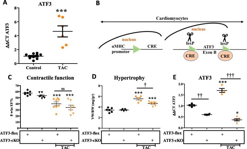
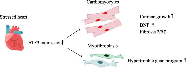
Background: Activating transcription 3 (ATF3) is a member of the basic leucine zipper family of transcription factors. ATF3 is an immediate early gene expressed following various cellular stresses. ATF3 acts through binding to cyclic AMP response elements found in the promoters of key regulatory proteins that determine cell fate. In the heart, multiple cardiac stresses result in chronic ATF3 expression. Transgenic mice with ATF3 expression in cardiomyocytes clearly demonstrate that ATF3 serves a leading role in heart hypertrophy, cardiac fibrosis, cardiac dysfunction and death. In contrast, the use of ATF3 whole body knockout mice resulted non-conclusive results. The heart is composed of various cell types such as cardiomyocytes, fibroblasts, endothelial and immune cells. The question that we addressed in this study is whether ablation of ATF3 in unique cell types in the heart results in diverse cardiac phenotypes.

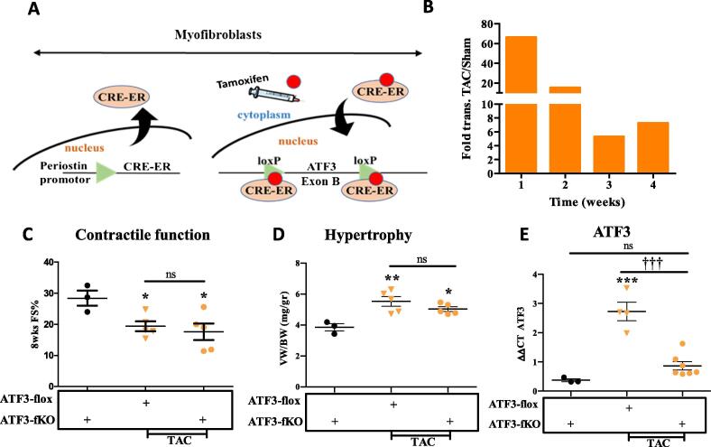
Methods: ATF3-flox mice were crossed with αMHC and Postn specific promoters directing CRE expression and thus ATF3 ablation in cardiomyocytes and myofibroblast cells. Mice were challenged with transverse aortic constriction (TAC) for eight weeks and heart function, ventricle weight, hypertrophic markers, fibrosis markers and ATF3 expression were assessed by qRT-PCR.

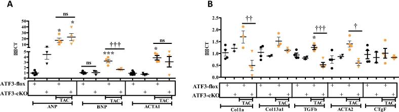
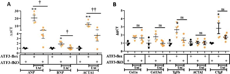
Results: The results of the study show that ATF3 deletion in cardiomyocytes followed by TAC resulted in reduced heart growth and dampened fibrosis response while ATF3 ablation in myofibroblasts displayed a reduced hypertrophic gene program.

Conclusions: TAC-operation results in increased ATF3 expression in both myofibroblasts and cardiomyocytes that promotes a hypertrophic program and fibrotic cardiac growth, respectively.

Keywords: Cardiac remodeling; Cardiomyocytes fibroblasts; Conditional knockout; Heart failure; Hypertrophy; Pressure overload.


Conflict of interest statement

The authors declare that they have no known competing financial interests or personal relationships that could have appeared to influence the work reported in this paper.

Figures

Fig. 1
Fig. 1
ATF3 expression in cardyomyocytes promotes heart growth following TAC operation. A. mRNA was extracted from ventricles and the expression level of ATF3 was analyzed by qRT-PCR normalized with HSP90 as housekeeping gene. ATF3 transcription at eight weeks following TAC and control. B. Schematic representation for cardiac specific KO. αMHC-CRE transgenic mouse strain was crossed with ATF3-flox mouse strain (ATF3-flox). CRE is expressed shortly before birth resulting in Exon B excision (loxP sites are indicated) specifically in cardiomyocytes. CRE expressing mice harboring two alleles of ATF3-flox are designated ATF3-cKO. C. Eight weeks following TAC procedure (10–12 weeks old male mice), mice were subjected to MRI. Ejection fraction (EF) was calculated as described in the methods. D. Mice were sacrificed eight weeks following TAC. The hearts were excised and the ventricles weight to body weight ratio is shown (VW/BW). E. mRNA was extracted from ventricles and the expression level of ATF3 was analyzed by qRT-PCR normalized with HSP90 as housekeeping gene.
Fig. 2
Fig. 2
Hypertrophic and Fibrosis gene markers in ATF3-cKO mice. mRNA was extracted from ventricles and the expression levels were measured by qRT-PCR normalized with HSP90 as housekeeping gene. A. Hypertrophic gene markers ANP, BNP and Acta1. B. Fibrosis gene markers Col1α1, Col3α1, TGFβ3, Acta2, CTGF were analyzed. Expression levels are presented as relative values (compared to ATF3-Flox control mice, defined as 1). All results represent the mean ± SE *** P ≤ 0.05, control vs. TAC; †P ≤ 0.05, difference between genotypes. Each dot or triangle represents one mouse. n.s. indicates no statistical difference.
Fig. 3
Fig. 3
ATF3 expression in myofibroblasts promotes hypertrophic gene program following TAC. A. Schematic representation for fibroblasts specific KO. Periostin-CRE-ER transgenic mouse strain was crossed with ATF3-flox mouse strain (ATF3-flox). CRE-ER is expressed in activated fibroblasts cells. Tamoxifen (red circle) injection is used to facilitate the translocation of CRE-ER into the nucleus resulting in Exon B excision (loxP sites are indicated) specifically in fibroblasts. CRE-ER expressing mice harboring two alleles of ATF3-flox are designated ATF3-fKO. B. RNA-sequencing data analysis for Periostin expression in the hearts of mice at different time points following TAC . C. Eight weeks following TAC (10–12 weeks old male mice), mice were subjected to echocardiography and fractional shortening was calculated as described in the methods. D. Mice were sacrificed eight weeks following TAC. The hearts were excised and the ventricles weight to body weight ratio is shown (VW/BW). E. mRNA was extracted from ventricles and the expression level of ATF3 was analyzed by qRT-PCR. (For interpretation of the references to colour in this figure legend, the reader is referred to the web version of this article.)
Fig. 4
Fig. 4
Hypertrophic and Fibrosis gene markers in ATF3-fKO mice. mRNA was extracted from ventricles and the expression level were measured by qRT-PCR. A. Hypertrophic gene markers ANP, BNP and Acta1 B. Fibrosis gene markers Col1α1, Col3α1, TGFβ3, Acta2, CTGF were analyzed. Expression levels are presented as relative values (compared to ATF3-Flox control mice, defined as 1). All results represent the mean ± SE *** P ≤ 0.05, control vs. TAC; †P ≤ 0.05, difference between genotypes. Each dot or triangle represents one mouse. n.s. indicates no statistical difference.
Fig. 5
Fig. 5
Schematic representation of the main conclusions. Pressure overload stress results in elevation of ATF3 expression in the heart. ATF3 in cardiomyocytes is responsible for cardiac growth and fibrosis gene markers while ATF3 expression in myofibroblasts promotes hypertrophic gene program.

Similar articles

Cited by

References

    1. Molkentin J.D., Dorn G.W., 2nd Cytoplasmic signaling pathways that regulate cardiac hypertrophy. Annu. Rev. Physiol. 2001;63:391–426. - PubMed
    1. Zou Y., Takano H., Akazawa H., Nagai T., Mizukami M., Komuro I. Molecular and cellular mechanisms of mechanical stress-induced cardiac hypertrophy. Endocr. J. 2002;49:1–13. - PubMed
    1. Bernardo B.C., Weeks K.L., Pretorius L., McMullen J.R. Molecular distinction between physiological and pathological cardiac hypertrophy: experimental findings and therapeutic strategies. Pharmacol. Ther. 2010;128:191–227. - PubMed
    1. Tardiff J.C. Cardiac hypertrophy: stressing out the heart. J. Clin. Investig. 2006;116:1467–1470. - PMC - PubMed
    1. Kehat I., Molkentin J.D. Molecular pathways underlying cardiac remodeling during pathophysiological stimulation. Circulation. 2010;122:2727–2735. - PMC - PubMed

LinkOut - more resources