Review
. 2021 Feb;57(2):191-206.
doi: 10.1007/s11626-020-00532-8. Epub 2021 Jan 12.

Advances in 3D neuronal microphysiological systems: towards a functional nervous system on a chip

Affiliations
Review

Advances in 3D neuronal microphysiological systems: towards a functional nervous system on a chip

Wesley A Anderson et al. In Vitro Cell Dev Biol Anim. 2021 Feb.

Abstract

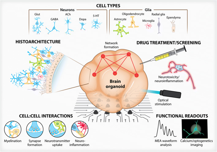
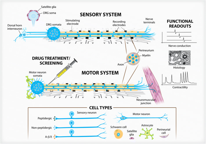
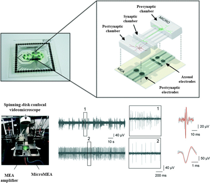
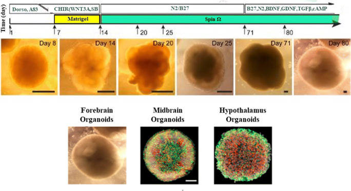
Microphysiological systems (MPS) designed to study the complexities of the peripheral and central nervous systems have made marked improvements over the years and have allowed researchers to assess in two and three dimensions the functional interconnectivity of neuronal tissues. The recent generation of brain organoids has further propelled the field into the nascent recapitulation of structural, functional, and effective connectivities which are found within the native human nervous system. Herein, we will review advances in culture methodologies, focused especially on those of human tissues, which seek to bridge the gap from 2D cultures to hierarchical and defined 3D MPS with the end goal of developing a robust nervous system-on-a-chip platform. These advances have far-reaching implications within basic science, pharmaceutical development, and translational medicine disciplines.

Keywords: 3D culture; Brain organoids; Microengineering; Microphysiological systems; Nerve-on-a-chip; Neuroscience.


Conflict of interest statement

WAA is a paid employee of AxoSim, Inc., New Orleans, LA, USA, which is a for-profit entity commercializing neural microphysiological system. TH and HH are inventors on a patent by Johns Hopkins University, which is licensed to AxoSim, for which they both consult and TH is a shareholder. MJM is an inventor on a patent by Tulane University also licensed by AxoSim, of which he is a co-founder and shareholder. AB declares no conflict of interest.

Figures

Figure 1.
Figure 1.
Three-dimensional brain organoid models. Advances in culture methodologies have allowed researchers to build complex, heterogenous brain organoids that are both structurally similar to in vivo architectures and provide meaningful functional readouts. Brain organoids, over traditional two-dimensional cultures, can integrate a myriad of neuronal and glial cell types which exhibit histoarchitecture and cell:cell interactions not possible on planar surfaces. Whether situated atop MEAs or engineered to contain optogenetic constructs, drug screening can be performed to assess brain organoid function and viability.
Figure 2.
Figure 2.
Three-dimensional peripheral nerve models. Peripheral nerve anatomy informs researchers to create 3D constructs that respect the compartmentalized and polar features found naturally. By containing soma either within restrictive chambers or growing them as spheroids, axon and glial outgrowth can be directed along MEA surfaces to assess electrophysiological function. Sensory neural constructs (top) can integrate a variety of neural subtypes and be used to study satellite glia in neuropathic pain models and the subsequent input into the dorsal horn. Motor neuron constructs (bottom) are currently being integrated into multiorgan NMJ models. Both systems are able to be myelinated by Schwann cells. Nerve fascicle formation, along with perineurial glia, can be studied.
Figure 3.
Figure 3.
MPS for modeling CNS interconnected functionality has been achieved with engineered culture systems using microfabricated chambers for specifying synaptic connectivity and microelectrodes for evaluating function. Adapted from Moutaux et al. (2018), with permission from The Royal Society of Chemistry.
Figure 4.
Figure 4.
Brain organoids rely on self-assembly during differentiation from pluripotent cells. Adjusting culture conditions has produced large organoids exhibiting self-organized internal structure as well as organoids representing distinct brain regions. Adapted from Qian et al. (2016), with permission from Elsevier.
Figure 5.
Figure 5.
Brain spheroids for the 3D modeling of the central nervous system. Developed spheroids show a diverse population of both (a) neuronal and (b) glial cells throughout the bulk of the organoid. (c) Myelination is also readily apparent and the progression of myelination within the spheroid can be assessed via both (d) immunohistochemistry and (e) TEM. Reproduced from Pamies et al. (2017), under Creative Commons license.
Figure 6.
Figure 6.
Peripheral nervous system compartmentalization through 3D printed constructs. (a) Schematic and formed 3D printed chamber for compartmentalized cell growth. From left to right, these three chambers contain (b) neuron cell bodies, (c) Schwann cells growing along axonal projections, and (d) axon terminals. Models such as this one can ensure compartmentalized growth and can realize aligned growth of various cell types. Adapted from Johnson et al. (2016), with permission from The Royal Society of Chemistry.
Figure 7.
Figure 7.
Peripheral nerve MPS demonstrating compartmentalization, polarization, and alignment in 2D, along with integration of microelectrodes for spike sorting and observation of axonal conduction. Adapted from Sakai et al. (2017), by permission of Oxford University Press.
Figure 8.
Figure 8.
Peripheral nerve modeling and myelination. (a) Rat primary sensory neuron tissue grown as a spheroid within a growth-restrictive hydrogel can yield (b) induced compound action potentials detected by extracellular field recording. (c, d) Similar spheroids composed of iPSC-derived human neurons can show outgrowth and myelination that can be confirmed by (e) TEM micrographs showing compact myelin rings. Reproduced from Sharma et al. (2019), under Creative Commons license.

References

    1. Abreu CM, Gama L, Krasemann S, Chesnut M, Odwin-Dacosta S, Hogberg HT, Hartung T, Pamies D. Microglia increase inflammatory responses in iPSC-derived human BrainSpheres. Front Microbiol. 2018;9:2766. doi: 10.3389/fmicb.2018.02766. - DOI - PMC - PubMed
    1. Afshar Bakooshli M, Lippmann ES, Mulcahy B, Iyer N, Nguyen CT, Tung K, et al. A 3D culture model of innervated human skeletal muscle enables studies of the adult neuromuscular junction. Elife. 2019;8:e44530. doi: 10.7554/eLife.44530. - DOI - PMC - PubMed
    1. Alepee N, Bahinski A, Daneshian M, De Wever B, Fritsche E, Goldberg A, et al. State-of-the-art of 3D cultures (organs-on-a-chip) in safety testing and pathophysiology. ALTEX. 2014;31(4):441–477. doi: 10.14573/altex.1406111. - DOI - PMC - PubMed
    1. Amin ND, Pasca SP. Building models of brain disorders with three-dimensional organoids. Neuron. 2018;100(2):389–405. doi: 10.1016/j.neuron.2018.10.007. - DOI - PubMed
    1. Anderson WA, Willenberg AR, Bosak AJ, Willenberg BJ, Lambert S. Use of a capillary alginate gel (Capgel) to study the three-dimensional development of sensory nerves reveals the formation of a rudimentary perineurium. J Neurosci Methods. 2018;305:46–53. doi: 10.1016/j.jneumeth.2018.05.003. - DOI - PubMed