

Intravascular ultrasound imaging in evaluation of aortic stiffness: A proof-of-concept study
- PMID: 33438178
- PMCID: PMC10129260
- DOI: 10.5603/CJ.a2021.0003
Intravascular ultrasound imaging in evaluation of aortic stiffness: A proof-of-concept study
Abstract
Background: Aortic stiffness is a well-known cardio-vascular risk factor. For years, different methods have been studied in the assessment of aortic elastic properties and large arterial stiffness for risk stratification. Herein is an assessment of the role of intravascular ultrasound (IVUS) imaging for the evaluation of aortic elastic properties.
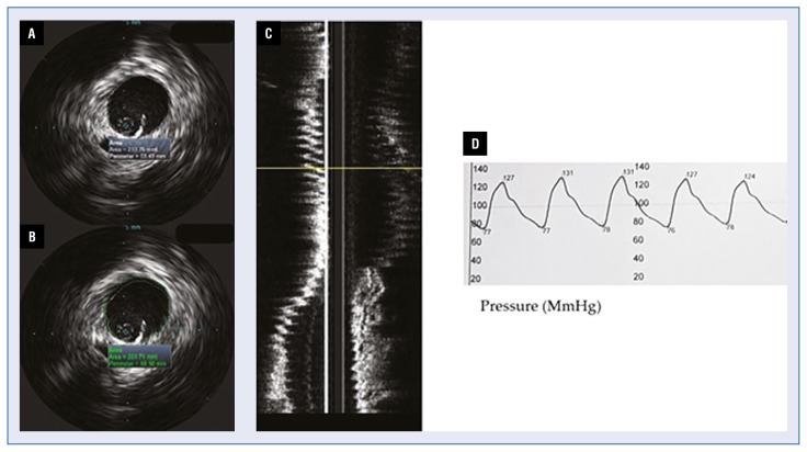
Methods: Intravascular ultrasound imaging of the aorta was performed in 12 patients with transthoracic echocardiography (TTE) and computed tomography (CT) evidence for enlargement of the ascending aorta - diameter ≥ 40.0 mm. Mechanical properties of the aorta were derived from the measured diameters and intra-aortic pressure. Paired samples T-test analyses were performed to determine differences between measurements derived by TTE, CT and IVUS.
Results: Mean values of the calculated elastic properties via IVUS of the ascending aorta were as follows: compliance 0.021 ± 0.02; strain 205 ± 4.3; aortic stiffness index 4.3 ± 0.75; elastic modulus 0.31 ± 0.05. On paired T-test analysis maximum ascending aortic diameter measured by CT aortography and IVUS did not differ significantly (t = -0.19, p = 0.985), but a significant difference between IVUS measurements and TTE derived diameters was found (t = 13.118, p = 0.034). On average, IVUS diameters were 2.3 mm larger than the results acquired by TTE (95% confidence interval: 14.21-17.13).
Conclusions: Intravascular ultrasound examination of the ascending aorta provided larger diameters than the ones collected by means of TTE. However, IVUS measurements did not differ significantly from diameters derived by CT aortography.
Keywords: aortic compliance; aortic stress; intravascular ultrasound.
Conflict of interest statement
Figures

Similar articles
-
Comparison of dynamic changes in aortic diameter during the cardiac cycle measured by computed tomography angiography and transthoracic echocardiography.J Vasc Surg. 2019 May;69(5):1538-1544. doi: 10.1016/j.jvs.2018.07.083. J Vasc Surg. 2019. PMID: 31010518
-
Differences in Aortic Diameter Measurements with Intravascular Ultrasound and Computed Tomography After Blunt Traumatic Aortic Injury.Ann Vasc Surg. 2018 Jul;50:148-153. doi: 10.1016/j.avsg.2017.11.056. Epub 2018 Feb 23. Ann Vasc Surg. 2018. PMID: 29481934
-
Comparison of intravascular ultrasound- and centerline computed tomography-determined aortic diameters during thoracic endovascular aortic repair.J Vasc Surg. 2017 Oct;66(4):1184-1191. doi: 10.1016/j.jvs.2017.03.445. Epub 2017 Jun 22. J Vasc Surg. 2017. PMID: 28648482
-
Accuracy of Intravascular Ultrasound Evaluation for the Assessment of Native Valve Measures in Patients Undergoing TAVI: Preliminary Results.Surg Technol Int. 2016 Oct 26;29:201-206. Surg Technol Int. 2016. PMID: 27466865
-
Ethnic Differences in Ascending Aorta Dimensions and Dilatation Rates: A Systematic Review.Cureus. 2024 Dec 31;16(12):e76703. doi: 10.7759/cureus.76703. eCollection 2024 Dec. Cureus. 2024. PMID: 39898145 Free PMC article. Review.
Cited by
-
Impact of renin-angiotensin system targeted therapy on aortic elastic properties assessed by computed tomography.Int J Cardiol Heart Vasc. 2024 Nov 22;55:101562. doi: 10.1016/j.ijcha.2024.101562. eCollection 2024 Dec. Int J Cardiol Heart Vasc. 2024. PMID: 39649025 Free PMC article.
References
MeSH terms
LinkOut - more resources
Full Text Sources
Other Literature Sources
