Review
. 2021 Feb 1;40(3):e104705.
doi: 10.15252/embj.2020104705. Epub 2021 Jan 13.

Molecular mechanisms and physiological functions of mitophagy

Affiliations
Review

Molecular mechanisms and physiological functions of mitophagy

Mashun Onishi et al. EMBO J. .

Abstract

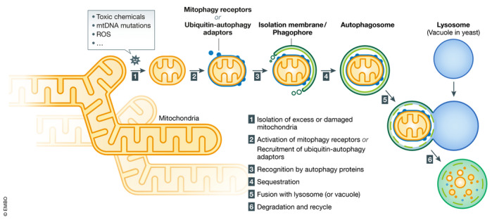
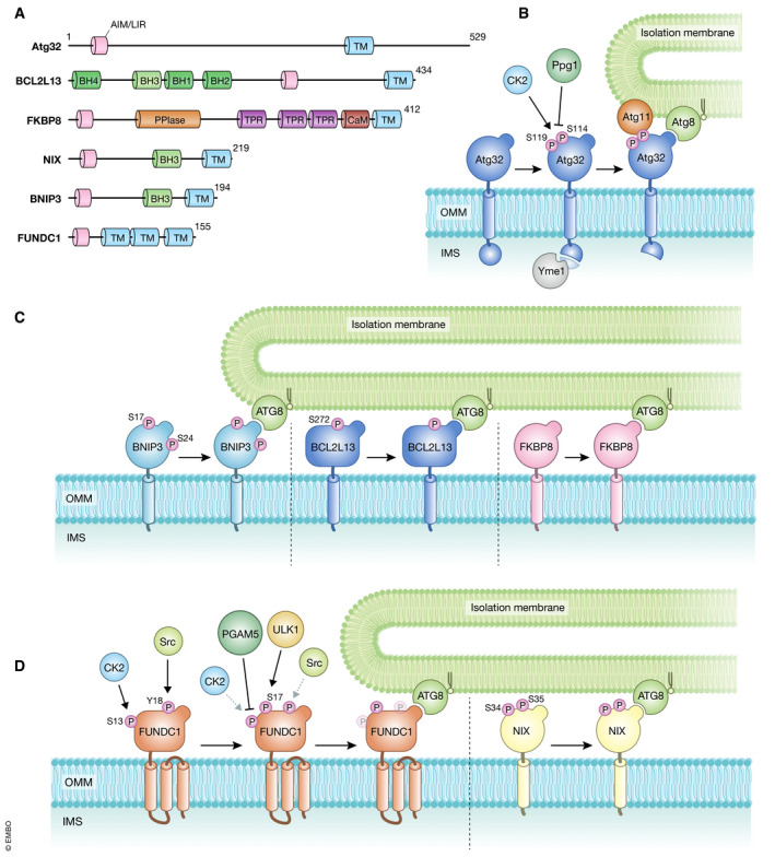
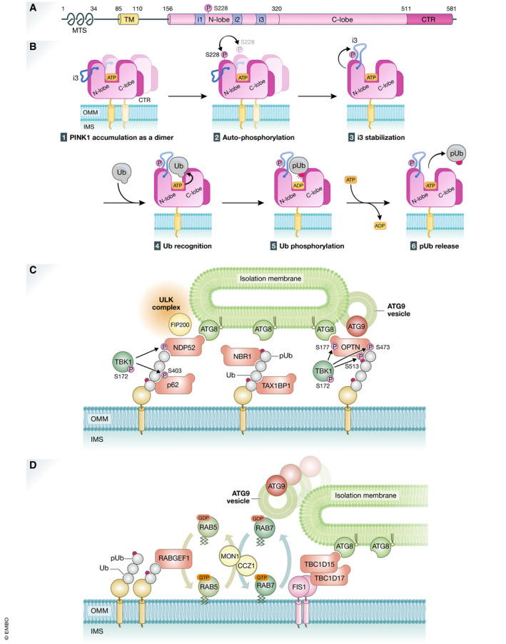
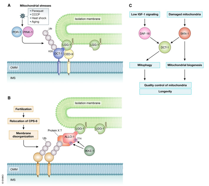
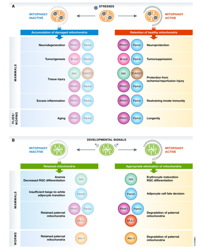
Degradation of mitochondria via a selective form of autophagy, named mitophagy, is a fundamental mechanism conserved from yeast to humans that regulates mitochondrial quality and quantity control. Mitophagy is promoted via specific mitochondrial outer membrane receptors, or ubiquitin molecules conjugated to proteins on the mitochondrial surface leading to the formation of autophagosomes surrounding mitochondria. Mitophagy-mediated elimination of mitochondria plays an important role in many processes including early embryonic development, cell differentiation, inflammation, and apoptosis. Recent advances in analyzing mitophagy in vivo also reveal high rates of steady-state mitochondrial turnover in diverse cell types, highlighting the intracellular housekeeping role of mitophagy. Defects in mitophagy are associated with various pathological conditions such as neurodegeneration, heart failure, cancer, and aging, further underscoring the biological relevance. Here, we review our current molecular understanding of mitophagy, and its physiological implications, and discuss how multiple mitophagy pathways coordinately modulate mitochondrial fitness and populations.

Keywords: autophagy; mitochondria; phosphorylation; quality and quantity control; ubiquitin.


Conflict of interest statement

The authors declare that they have no conflict of interest.

Figures

Figure 1
Figure 1. Overview of mitophagy
(1) Intra‐ and extracellular cues promote isolation of excess or damaged mitochondria via fragmentation of tubular networks. (2) Mitophagy receptors or ubiquitin–autophagy adaptors that confer selectivity for degradation are recruited and/or activated on the surface of mitochondria. (3) Core autophagy‐related proteins target to mitochondria and generate the isolation membrane/phagophore surrounding mitochondria. (4) Targeted mitochondria are enclosed and sequestrated by autophagosomes. (5) Autophagosomes are transported and fused with lytic compartments such as vacuoles in yeast or lysosomes in mammals. (6) Lysosomal or vacuolar acidic hydrolases flow into autophagosomes to degrade mitochondria, and the contents will be recycled.
Figure 2
Figure 2. Receptor‐mediated mitophagy
(A) Schematic representation of the domain structures of mitophagy receptors in yeast and mammals. AIM/LIR, Atg8‐family protein‐interacting motif/LC3‐interacting region (pink); TM, transmembrane domain (light blue); BH1‐4, Bcl‐2 homology 1‐4 domain (green and light green); PPlase, peptidyl‐prolyl cis‐trans isomerase domain (orange); TPR, tetratricopeptide repeat domain (purple); CaM, calmodulin‐binding domain (dark red). The protein size is indicated as the number of amino acids. (B‐D) Models for mitophagy receptor activation and protein recruitment on the mitochondrial surface. The yeast mitophagy receptor Atg32 (B), and the mammalian mitophagy receptors BNIP3, BCL2L13, FKBP8 (C), FUNDC1, and NIX (D) bind to ATG8 family proteins and then target the autophagy machinery to mitochondria. Phosphorylation and dephosphorylation serve as regulatory mechanisms to modulate the activity of mitophagy receptors. For details, see text.
Figure 3
Figure 3. PINK1 and ubiquitin‐mediated mitophagy
(A) Schematic depiction of the domain structures of PINK1. The protein and domain sizes are indicated as the number of amino acids. MTS, mitochondrial targeting signal; TM, transmembrane segment; N‐lobe and C‐lobe, N‐terminal and C‐terminal lobes found in a typical kinase, respectively. i1, i2, and i3, the insert regions unique to PINK1; CTR, C‐terminal region conserved among PINK1 homologs. (B) Molecular mechanisms underlying ubiquitin phosphorylation by PINK1 on depolarized mitochondria. (1) PINK1 forms a dimer on damaged mitochondria. (2) Ser228 is phosphorylated via intermolecular autophosphorylation in dimerized PINK1. (3) Ser228 phosphorylation stabilizes and underpins “insert 3 (i3)” at the correct position. (4) Ubiquitin (Ub) is recognized by PINK1 as a genuine substrate, (5) Ub Ser65 residue is phosphorylated via ATP hydrolysis, and (6) phosphorylated Ub (pUb) is released. (C) Recruitment of the core autophagy proteins and isolation membranes to mitochondria during PINK1/Parkin‐mediated mitophagy. Poly‐ubiquitin chains on damaged mitochondria are recognized directly by various autophagy adaptors. They are phosphorylated by TBK1 kinase, and the phosphorylation enhances the binding affinity to ubiquitin chains and ATG8 family proteins. NDP52 and OPTN specifically recruit ULK complex via FIP200 and ATG9 vesicles, respectively. (D) RABGEF1 recruited to mitochondria by poly‐ubiquitin chains triggers endosomal Rab cycles including RAB5 and the MON1/CCZ1 complex. MON1/CCZ1 directs RAB7 to mitochondria, and RAB7 facilitates the assembly of ATG9 vesicles to the autophagosome formation site. Mitochondrial Rab‐GAPs, TBC1D15 and TBC1D17, assist to complete RAB7 cycles and interact with ATG8 family proteins to recruit the isolation membrane.
Figure 4
Figure 4. Regulation of mitophagy in C. elegans
(A) PINK‐1/PDR‐1‐mediated mitophagy in somatic cells. DCT‐1 functions as an autophagy adaptor in association with CED‐9. DCT‐1 Lys26 (K26) is ubiquitinated in a PINK‐1/PDR‐1‐dependent manner. (B) Mechanism of allophagy in embryos. Fertilization triggers relocation of CPS‐6, the mitochondrial endonuclease, leading to membrane disorganization and ubiquitylation of paternal mitochondria. Ubiquitin (Ub) molecules on paternal mitochondria are recognized directly or indirectly by the autophagy adaptor ALLO‐1. IKKE‐1‐dependent phosphorylation of ALLO‐1 is also important for allophagy. (C) Transcriptional regulation of DCT‐1 contributes to mitochondrial homeostasis and longevity.
Figure 5
Figure 5. Physiological functions of mitophagy against mitochondrial stresses
(A) Mitochondria are constantly challenged by a subset of mitochondrial stresses such as oxidative stress. Mitophagy contributes to mitochondrial quality control and prevention of pathologies including neurodegeneration, tumorigenesis, tissue injury, excess inflammation, and aging. (B) Physiological functions of mitophagy during development and differentiation. Mitochondrial elimination in response to developmental cues is crucial for maturation of cells and tissues.

References

    1. Agnihotri S, Golbourn B, Huang X, Remke M, Younger S, Cairns RA, Chalil A, Smith CA, Krumholtz SL, Mackenzie D et al (2016) PINK1 is a negative regulator of growth and the Warburg effect in glioblastoma. Cancer Res 76: 4708–4719 - PubMed
    1. Al Rawi S, Louvet‐Vallee S, Djeddi A, Sachse M, Culetto E, Hajjar C, Boyd L, Legouis R, Galy V (2011) Postfertilization autophagy of sperm organelles prevents paternal mitochondrial DNA transmission. Science 334: 1144–1147 - PubMed
    1. Al Rawi S, Louvet‐Vallee S, Djeddi A, Sachse M, Culetto E, Hajjar C, Boyd L, Legouis R, Galy V (2012) Allophagy: a macroautophagic process degrading spermatozoid‐inherited organelles. Autophagy 8: 421–423 - PMC - PubMed
    1. Allen GF, Toth R, James J, Ganley IG (2013) Loss of iron triggers PINK1/Parkin‐independent mitophagy. EMBO Rep 14: 1127–1135 - PMC - PubMed
    1. Altshuler‐Keylin S, Shinoda K, Hasegawa Y, Ikeda K, Hong H, Kang Q, Yang Y, Perera RM, Debnath J, Kajimura S (2016) Beige adipocyte maintenance is regulated by autophagy‐induced mitochondrial clearance. Cell Metab 24: 402–419 - PMC - PubMed

Publication types

Substances

LinkOut - more resources