. 2021 Jan 13;17(1):e1009215.
doi: 10.1371/journal.ppat.1009215. eCollection 2021 Jan.

Injection site vaccinology of a recombinant vaccinia-based vector reveals diverse innate immune signatures

Affiliations

Injection site vaccinology of a recombinant vaccinia-based vector reveals diverse innate immune signatures

Jessamine E Hazlewood et al. PLoS Pathog. .

Abstract

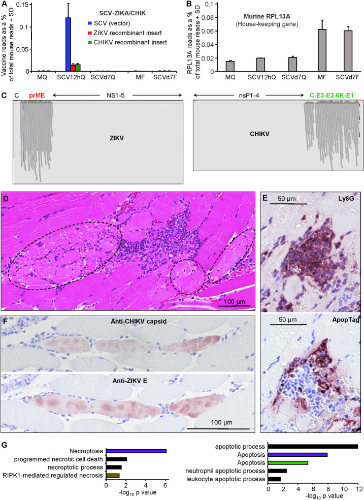
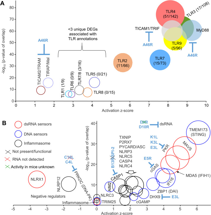
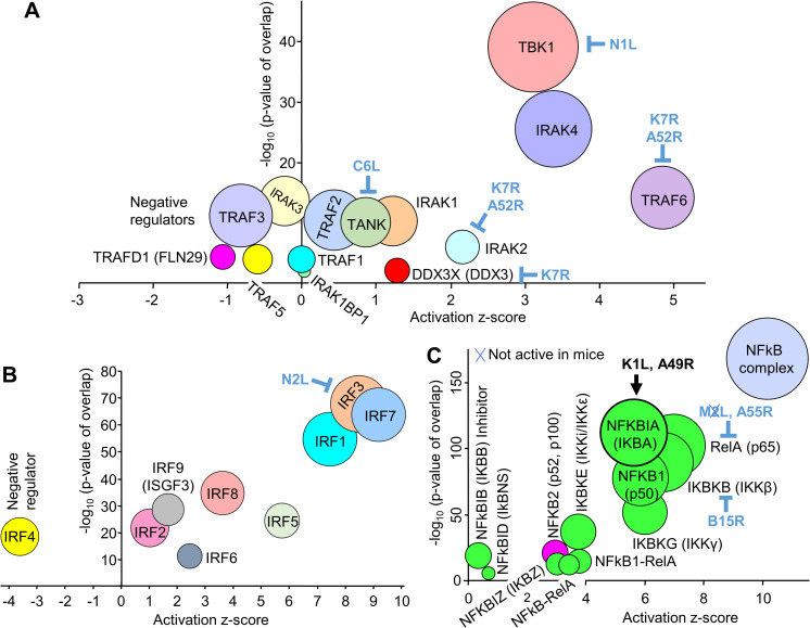
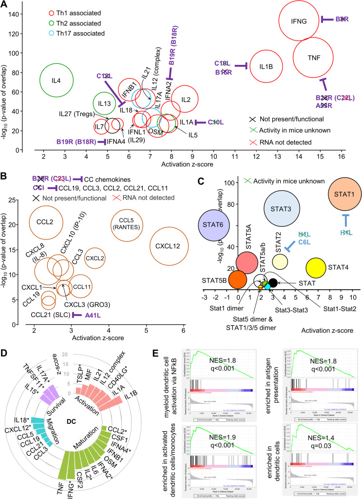
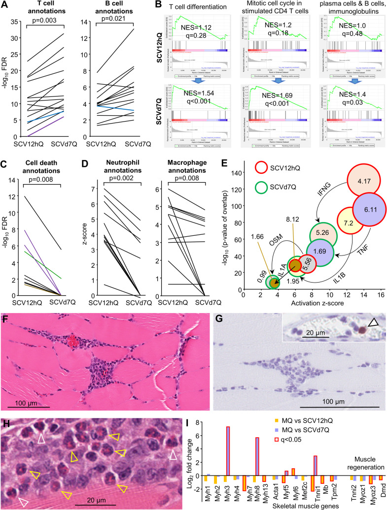
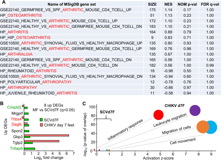
Poxvirus systems have been extensively used as vaccine vectors. Herein a RNA-Seq analysis of intramuscular injection sites provided detailed insights into host innate immune responses, as well as expression of vector and recombinant immunogen genes, after vaccination with a new multiplication defective, vaccinia-based vector, Sementis Copenhagen Vector. Chikungunya and Zika virus immunogen mRNA and protein expression was associated with necrosing skeletal muscle cells surrounded by mixed cellular infiltrates. The multiple adjuvant signatures at 12 hours post-vaccination were dominated by TLR3, 4 and 9, STING, MAVS, PKR and the inflammasome. Th1 cytokine signatures were dominated by IFNγ, TNF and IL1β, and chemokine signatures by CCL5 and CXCL12. Multiple signatures associated with dendritic cell stimulation were evident. By day seven, vaccine transcripts were absent, and cell death, neutrophil, macrophage and inflammation annotations had abated. No compelling arthritis signatures were identified. Such injection site vaccinology approaches should inform refinements in poxvirus-based vector design.


Conflict of interest statement

I have read the journal's policy and the authors of this manuscript have the following competing interests: NAP, LL and JDH own Sementis shares. JDH is the current CSO of Sementis. AS was a consultant for Sementis. PMH was the previous CEO/CSO of Sementis. LL and NAP have had, and/or currently have, salary and/or project support from funds provided, whole or in part, via Sementis. Sementis had no role in the design and interpretation of the study, or in preparation of the manuscript.

Figures

Fig 1
Fig 1. Vaccine genome read alignments, histology and immunohistochemistry.
(A) RNA-Seq reads from each of the five groups aligned to the three viral genomes (the vector, SCV, and the two recombinant immunogen inserts from ZIKV and CHIKV); MQ—quadriceps muscles from mock vaccinated, SCV12hQ mice quadriceps muscles from SCV-ZIKA/CHIK vaccinated mice 12 hours post vaccination, SCVd7Q -quadriceps muscles from SCV-ZIKA/CHIK vaccinated mice taken 7 days post vaccination, MF–feet from mock vaccinated mice 7 days post vaccination, and SCVd7F - feet from SCV-ZIKA/CHIK vaccinated mice 7 days post vaccination. The number of viral reads is expressed as a percentage of the number of reads mapping to the mouse genome, with 3 biological replicates providing the SD (S1 Fig). The bars plotting to ≈0% had values ranging from 0 to 3.5x10⁻⁵%. (B) RNA-Seq reads from each of the five groups aligned to the house-keeping gene, RPL13A, also expressed as a percentage of the number of reads mapping to the mouse genome. (C) IGV visualization of reads aligned to the recombinant structural polyprotein immunogens of ZIKV (prME) and CHIKV (C-E3-E2-6K-E1), which are encoded in the SCV-ZIKA/CHIK vaccine. All reads from all replicates are shown (for details see (S1A Table). As expected, no reads mapped to the non-structural genes of ZIKV or CHIKV (NS1-5 and nsP1-4, respectively), as these are not encoded in SCV-ZIKA/CHIK. (Vertical purple lines for ZIKV indicate base call errors after a string of Gs). (Reads mapping to the SCV genome are shown in S1B Table). (D) H&E staining of injection site 12 hours post vaccination. Dotted ovals indicate muscle cells in early stages of necrosis (pink staining). (E) Top; IHC with anti-Ly6G staining for neutrophils (parallel section to D focusing on area of infiltrates). Bottom: Apoptag staining of the same area, illustrating apoptosis within areas of infiltrating cells. (F) Top: IHC for CHIK capsid protein. Bottom: parallel section showing IHC for ZIKA E protein. (G) Cell death annotation from Cytoscape analysis of up-regulated DEGs at 12 hours post vaccination (MQ vs SCV12hQ) (S2C Table) divided into non-apoptotic signatures (left) and apoptotic signatures (right). KEGG Pathways (purple), Go process (black), Reactome Pathways (brown), UniProt Keywords (green).
Fig 2
Fig 2. TLR and cytosolic sensor signatures at 12 hours post vaccination.
(A) TLR signatures identified by IPA USR analysis (S2D Table) of 1608 DEGs identified in quadriceps muscles 12 hours post SCV-ZIKA/CHIK vaccination (MQ vs SCV12hQ; S2B Table). Circle diameters reflect the number of DEGs associated with each IPA USR annotation. Numbers in brackets indicate the number of unique DEGs associated with each annotation over the total number of DEGs associated with the TLR annotation; circles with colored fills contain >3 DEGs uniquely associated with the indicated TLR annotation. A46R is expressed in the cytoplasm of infected cells. (B) Cytosolic sensor signatures identified by IPA USR analysis (S2D Table). Sensors divided into 3 categories associated with dsRNA (red circles), DNA (blue circles) and inflammasome activation (black circles). Circle diameters reflect the number of DEGs associated with each annotation. VACV genes encoding cytoplasmic inhibitors are shown in blue, with the black cross indicating that the gene/protein is not present or not functional in SCV (or in the Copenhagen strain of VACV), the red cross indicating that the gene was not detected by RNA-Seq of MQ vs SCV12hQ, the green cross indicate that the activity in mice is unknown (see S1B Table).
Fig 3
Fig 3. Secondary messenger signatures at 12 hours post vaccination.
(A) Secondary messenger signatures. VACV encodes a series of cytoplasmic inhibitors, which are indicated in blue text. (B) Interferon response factors (IRFs). (C) NF-κB signatures. Green fill indicates canonical pathways, magenta fill non-canonical pathway, blue fill—not assigned to canonical or non-canonical. VACV-encoded cytoplasmic inhibitors are shown in blue text. K1L and A49R (black text) enhance the activity of NFKBIA, an inhibitor of the canonical pathway. Blue cross means the inhibitor is not active in mice.
Fig 4
Fig 4. Cytokine, chemokine, dendritic cell and STAT signatures at 12 hours post vaccination.
(A) Cytokine signatures. VACV genes encoding secreted inhibitors are shown in purple text. Black crosses indicate the inhibitors are not present or functional in SCV. Green crosses indicate that the activity in mice is unknown. Red crosses indicate the RNA was not detected in our RNA-Seq analysis. (B) Chemokine signatures. Purple text and crosses as in A. (C) STAT signatures. VACV encoded cytoplasmic inhibitors are shown in blue text. Crosses as in A. (D) IPA USRs associated with stimulation of dendritic cells. * indicates that the mediators have more than one of the four dendritic activities indicated. (The figure includes some USRs present in previous bubble graphs). For references see S2F Table. (E) GSEAs for the Blood Transcript Modules (right to left) M43.0 and M43.1 (gene sets combined, n = 21); M95.0, M95.1, M71 and M200 (gene sets combined, n = 49); M64, M67, M119 and M165) (gene sets combined, n = 71); M168 (n = 19). For gene set details see S2F Table.
Fig 5
Fig 5. The injection site day 7 post vaccination.
(A) Cytoscape analysis of up-regulated DEGs from MQ vs SCV12hQ and MQ vs SCVd7Q, illustrating the upward trend in significance of top B and T cell associated annotations. Black line–GO Process, Blue line GO—Component, Purple line—KEGG Pathways. For full lists and descriptions of annotations see S2J Table; statistics by paired t test for the full lists (parametric data distribution). (B) GSEAs were used to interrogate MQ vs SCV12hQ and MQ vs SCVd7Q gene lists using T cell and B cell BTMs (left to right) M14 (n = 12); M4.5 (n = 35); M156.0 and M156.1 (gene sets combined, n = 56) (for details of BTM gene sets see S2J Table). (C) As for A illustrating the downward trend of cell death annotations. Anotations not identified by the IPA analysis were nominally given a -log10 FDR value of zero (y axis). Color coding as for A, but also Green line–UniProt Keywords, Brown line–Reactome Pathways. For descriptions of annotations see S2K Table. (Statistics by Wilcoxon Signed Rank tests; non-parametric data distribution). (D) IPA Diseases and Functions analysis of DEGs (up and down-regulated) from MQ vs SCV12hQ and MQ vs SCVd7Q, illustrating the downward trend in z-scores for macrophage and neutrophil annotations (for description of annotations see S2L Table and S2M Table). (Statistics by Wilcoxon Signed Rank tests). (E) Major IPA USR pro-inflammatory cytokine annotations identified at 12 hours (Fig 4A and S2D Table) had much lower z-scores and p values on day 7 post vaccination (S2N Table). Numbers in the circles represent the log2 fold change for that cytokine relative to MQ. (F) H&E staining of intramuscular injection site lesions on day 7 post infection. (G) Neutrophil Ly6G staining of lesions from day 7 post infection. Arrow in insert shows positive staining of a neutrophil in a blood vessel capillary. (H) Eosinophils in the intramuscular injection site lesions on day 7 post vaccination. White arrow heads—mature segmented eosinophils. Yellow arrowheads–immature band eosinophils. (I) Expression of skeletal muscle genes from MQ vs SCV12hQ (S2A Table) and MQ vs SCVd7Q (S2G Table); bars with red outline indicate significant fold change (q<0.05).
Fig 6
Fig 6. No compelling arthritic signatures after SCV vaccination.
(A) MSigDB contains 16 gene sets associated with arthritis/arthralgia. These gene sets were used in GSEA analyses against the complete pre-ranked gene list for MF vs SCVd7F (SP2 Table). (B) MF vs SCVd7F provided 22 DEGs (with 2 filters applied q<0.05 and count sum >6) of which 8 were up-regulated (Table S2Q). Three of these were also up-regulated DEGs for CHIKV arthritis day 7 post infection (Table S2O; q<0.05). Red text–gene products associated with pro-inflammation activities. Green text–gene products with anti-inflammatory activities. (C) The 22 DEGs analyzed by IPA Diseases and Functions feature (direct and indirect) (S2Q Table) and compared with the same annotations identified by IPA analysis of DEGs for CHIKV arthritis (S2O Table).

Similar articles

Cited by

References

    1. Prow NA, Jimenez Martinez R, Hayball JD, Howley PM, Suhrbier A. Poxvirus-based vector systems and the potential for multi-valent and multi-pathogen vaccines. Expert Rev Vaccines. 2018;17: 925–934. 10.1080/14760584.2018.1522255 - DOI - PubMed
    1. Koch T, Dahlke C, Fathi A, Kupke A, Krahling V, Okba NMA, et al. Safety and immunogenicity of a modified vaccinia virus Ankara vector vaccine candidate for Middle East respiratory syndrome: an open-label, phase 1 trial. Lancet Infect Dis. 2020;20: 827–838. 10.1016/S1473-3099(20)30248-6 - DOI - PMC - PubMed
    1. Sutter G. A vital gene for modified vaccinia virus Ankara replication in human cells. Proc Natl Acad Sci U S A. 2020;117: 6289–6291. 10.1073/pnas.2001335117 - DOI - PMC - PubMed
    1. Pantaleo G, Janes H, Karuna S, Grant S, Ouedraogo GL, Allen M, et al. Safety and immunogenicity of a multivalent HIV vaccine comprising envelope protein with either DNA or NYVAC vectors (HVTN 096): a phase 1b, double-blind, placebo-controlled trial. Lancet HIV. 2019;6: e737–e749. 10.1016/S2352-3018(19)30262-0 - DOI - PMC - PubMed
    1. Laher F, Moodie Z, Cohen KW, Grunenberg N, Bekker LG, Allen M, et al. Safety and immune responses after a 12-month booster in healthy HIV-uninfected adults in HVTN 100 in South Africa: A randomized double-blind placebo-controlled trial of ALVAC-HIV (vCP2438) and bivalent subtype C gp120/MF59 vaccines. PLoS Med. 2020;17: e1003038 10.1371/journal.pmed.1003038 - DOI - PMC - PubMed

Publication types

MeSH terms

Substances