. 2021 Jan 12;16(1):20-28.
doi: 10.1016/j.stemcr.2020.11.017.

Developmental Stage-Specific Changes in Protein Synthesis Differentially Sensitize Hematopoietic Stem Cells and Erythroid Progenitors to Impaired Ribosome Biogenesis

Affiliations

Developmental Stage-Specific Changes in Protein Synthesis Differentially Sensitize Hematopoietic Stem Cells and Erythroid Progenitors to Impaired Ribosome Biogenesis

Jeffrey A Magee et al. Stem Cell Reports. .

Abstract

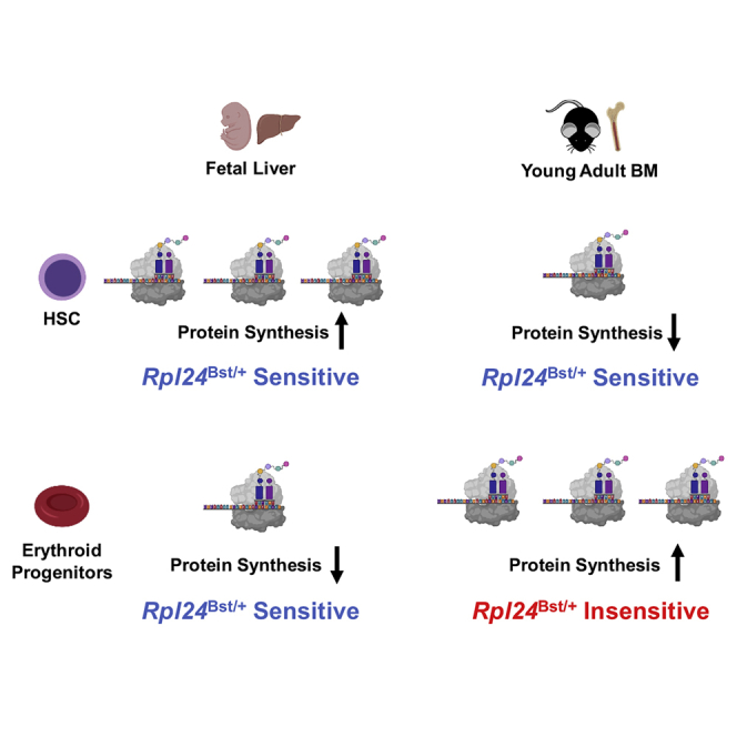
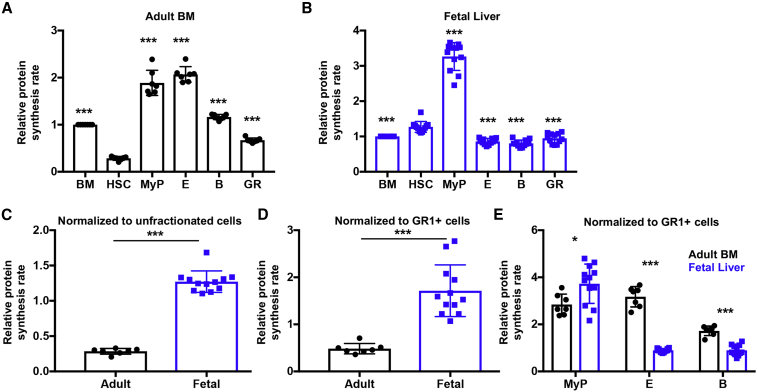
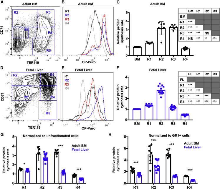
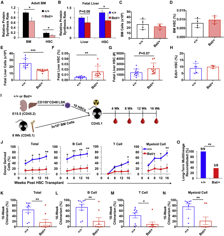
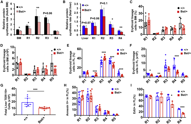
Adult hematopoietic stem cell (HSC) self-renewal requires precise control of protein synthesis, but fetal and adult HSCs have distinct self-renewal mechanisms and lineage outputs. This raises the question of whether protein synthesis rates change with age. Here, we show that protein synthesis rates decline during HSC ontogeny, yet erythroid protein synthesis rates increase. A ribosomal mutation that impairs ribosome biogenesis (Rpl24Bst/+) disrupts both fetal and adult HSC self-renewal. However, the Rpl24Bst/+ mutation selectively impairs fetal erythropoiesis at differentiation stages that exhibit fetal-specific attenuation of protein synthesis. Developmental changes in protein synthesis thus differentially sensitize hematopoietic stem and progenitor cells to impaired ribosome biogenesis.

Keywords: erythroid; erythropoiesis; hematopoiesis; hematopoietic stem cell; progenitor; protein synthesis; proteostasis; ribosome; ribosomopathy; translation.


Figures

None
Graphical abstract
Figure 1
Figure 1
Distinct Protein Synthesis Rates in Fetal and Adult Hematopoietic Stem and Progenitor Cells (A and B) Protein synthesis in hematopoietic stem and progenitor cells relative to unfractionated cells in (A) young adult BM and (B) E15.5 fetal liver. Data are shown for unfractionated BM cells, liver cells, CD150+CD48LSK HSCs, CD127LineageSCA1CD117+ myeloid progenitors (MyP), CD71+TER119+ erythroid progenitors (E), IgMB220+ B lineage progenitors (B), GR1+ cells (GR). (C and D) Relative protein synthesis in young adult BM and fetal liver HSCs normalized to (C) unfractionated cells or (D) GR1+ cells. (E) Relative protein synthesis in young adult BM and fetal liver restricted hematopoietic progenitor cells normalized to GR1+ cells. All data represent mean ± SD. N = 7 adult mice and 11 embryos. Statistical significance was assessed relative to HSCs using a repeated-measures one-way ANOVA followed by Dunnett's multiple comparisons test (A and B), or using a two-tailed Student's t test to compare differences between fetal and adult cells (C–E); p < 0.05, ∗∗p < 0.01, ∗∗∗p < 0.001.
Figure 2
Figure 2
Dynamic Changes in Protein Synthesis during Erythroid Differentiation Are Distinct in Adult Bone Marrow and Fetal Liver (A) Representative flow cytometry plot showing gating strategy for R1-R5 erythroid lineage cells in young adult BM. (B) Representative histograms showing OP-Puro incorporation in R1-R4 erythroid progenitors in young adult BM. (C) Relative protein synthesis based on OP-Puro incorporation in young adult R1-R4 erythroid progenitors relative to unfractionated BM cells in vivo. Statistical differences are summarized in the adjacent table. (D) Representative flow cytometry plot showing gating strategy for R1-R5 erythroid lineage cells in E15.5 fetal liver. (E) Representative histograms showing OP-Puro incorporation in R1-R4 erythroid progenitors in E15.5 fetal liver. (F) Relative protein synthesis based on OP-Puro incorporation in E15.5 fetal liver (FL) R1-R4 erythroid progenitors relative to unfractionated liver cells in vivo. Statistical differences are summarized in the adjacent table. (G and H) Relative protein synthesis in young adult BM and E15.5 fetal liver R1-R4 erythroid progenitors normalized to (G) unfractionated cells or (H) GR1+ cells. All data represent mean ± SD. N = 7 adult mice and 11 embryos. Statistical significance was assessed using a repeated-measures one-way ANOVA followed by Tukey's multiple comparisons test (C and F), or using a two-tailed Student's t test to compare differences between fetal and adult cells (G and H); p < 0.05, ∗∗p < 0.01, ∗∗∗p < 0.001.
Figure 3
Figure 3
Fetal Liver Rpl24Bst/+ HSCs Have Impaired Long-Term Multilineage Reconstituting Activity (A and B) Relative protein synthesis rates in (A) unfractionated BM and HSCs in young adult Rpl24Bst/+ and control mice, and (B) unfractionated fetal liver cells and HSCs in E15.5 Rpl24Bst/+ and littermate control embryos. Values are normalized to control BM (N = 4 mice/genotype) or control liver cells (N = 12–13 embryos/genotype). (C and D) (C) BM cellularity and (D) HSC frequency in young adult Rpl24Bst/+ and control mice (1 femur +1 tibia/mouse; N = 4 mice/genotype). (E–G) (E) Fetal liver cellularity, (F) HSC frequency, and (G) HSC number in E15.5 Rpl24Bst/+ and littermate control embryos (N = 10 embryos/genotype). (H) Frequency of fetal liver HSCs that incorporated EdU after a 1-h pulse in vivo (N = 3–5 embryos/genotype). (I) Diagram of experimental strategy to test long-term multilineage reconstituting activity of fetal liver Rpl24Bst/+ HSCs. (J) Donor-cell engraftment when 10 Rpl24Bst/+ (Bst/+) or littermate control (+/+) HSCs were transplanted with 3 × 105 recipient-type young adult BM cells into irradiated mice. Total hematopoietic, B-, T-, and myeloid-cell engraftment is shown 4, 8, 12, and 16 weeks after transplantation (N = 8–9 recipients per genotype). (K–N) Long-term (16-week) donor (K) hematopoietic, (L) B, (M) T, and (N) myeloid cell engraftment in the peripheral blood of individual recipient mice from (J). (O) Frequency of recipient mice in (J) that exhibited long-term (16-week) multilineage reconstitution (≥0.5% donor-derived peripheral blood B, T, and myeloid cells). Data represent mean ± SD (A–H) or SEM (J–N). Statistical significance was assessed using a two-tailed Student's t test or a Fisher's exact test (O); p < 0.05, ∗∗p < 0.01, ∗∗∗p < 0.001.
Figure 4
Figure 4
The Rpl24Bst/+ Mutation Impairs Fetal But Not Adult Erythroid Lineage Cells (A and B) Relative protein synthesis in Rpl24Bst/+ (Bst/+) or control (+/+) erythroid progenitor cells in (A) young adult BM (N = 4 mice/genotype) and (B) E15.5 fetal liver (N = 12–13 embryos/genotype) in vivo. (C) Frequency and (D) absolute number of erythroid progenitors in Rpl24Bst/+ (Bst/+) or control (+/+) young adult BM (1 femur +1 tibia/mouse; N = 3–4 mice/genotype). (E) Frequency and (F) absolute number of erythroid progenitors in Rpl24Bst/+ (Bst/+) or control (+/+) E15.5 fetal liver (FL; N = 10 embryos/genotype). (G) Number of TER119+ cells in Rpl24Bst/+ (Bst/+) or control (+/+) E15.5 fetal liver (N = 10 embryos/genotype). (H) Frequency of erythroid progenitors that are Annexin V+ in Rpl24Bst/+ (Bst/+) or control (+/+) E15.5 fetal liver (N = 7 embryos/genotype). (I) Frequency of Rpl24Bst/+ (Bst/+) or control (+/+) fetal liver erythroid progenitors that incorporated EdU after a 1-h pulse in vivo (N = 3–5 embryos/genotype). Data represent mean ± SD. Statistical significance was assessed using a two-tailed Student's t test; p < 0.05, ∗∗p < 0.01, ∗∗∗p < 0.001.

Similar articles

Cited by

References

    1. Akashi K., Traver D., Miyamoto T., Weissman I.L. A clonogenic common myeloid progenitor that gives rise to all myeloid lineages. Nature. 2000;404:193–197. - PubMed
    1. Armistead J., Triggs-Raine B. Diverse diseases from a ubiquitous process: the ribosomopathy paradox. FEBS Lett. 2014;588:1491–1500. - PubMed
    1. Basak A., Munschauer M., Lareau C.A., Montbleau K.E., Ulirsch J.C., Hartigan C.R., Schenone M., Lian J., Wang Y., Huang Y. Control of human hemoglobin switching by LIN28B-mediated regulation of BCL11A translation. Nat. Genet. 2020;52:138–145. - PMC - PubMed
    1. Cai X., Gao L., Teng L., Ge J., Oo Z.M., Kumar A.R., Gilliland D.G., Mason P.J., Tan K., Speck N.A. Runx1 deficiency decreases ribosome biogenesis and confers stress resistance to hematopoietic stem and progenitor cells. Cell Stem Cell. 2015;17:165–177. - PMC - PubMed
    1. Chua B.A., Signer R.A.J. Hematopoietic stem cell regulation by the proteostasis network. Curr. Opin. Hematol. 2020;27:254–263. - PMC - PubMed

Publication types

MeSH terms