. 2021 Mar:204:108436.
doi: 10.1016/j.exer.2021.108436. Epub 2021 Jan 10.

Knockdown of TRIM5α or TRIM11 increases lentiviral vector transduction efficiency of human Muller cells

Affiliations

Knockdown of TRIM5α or TRIM11 increases lentiviral vector transduction efficiency of human Muller cells

Monica M Sauter et al. Exp Eye Res. 2021 Mar.

Abstract

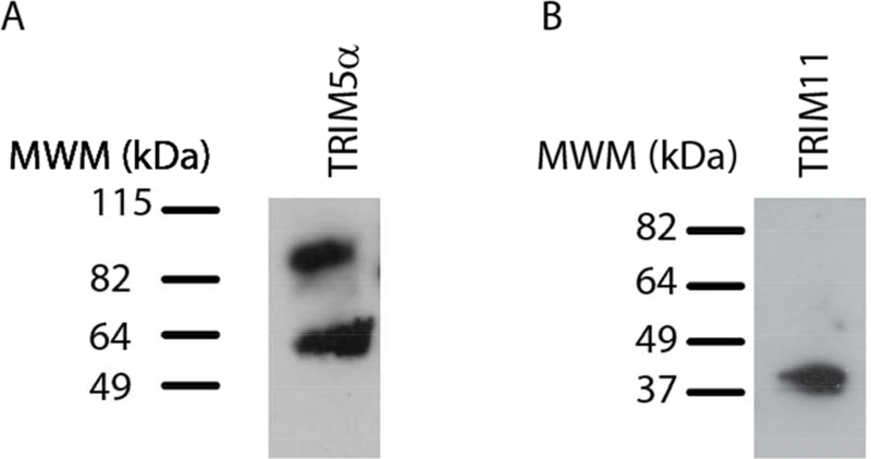
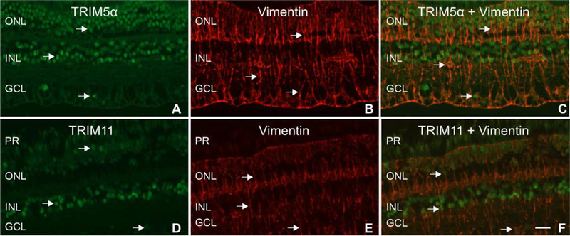
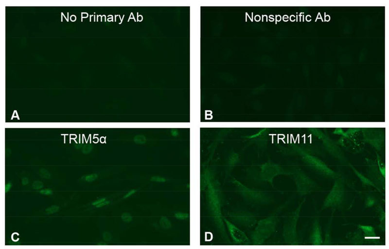
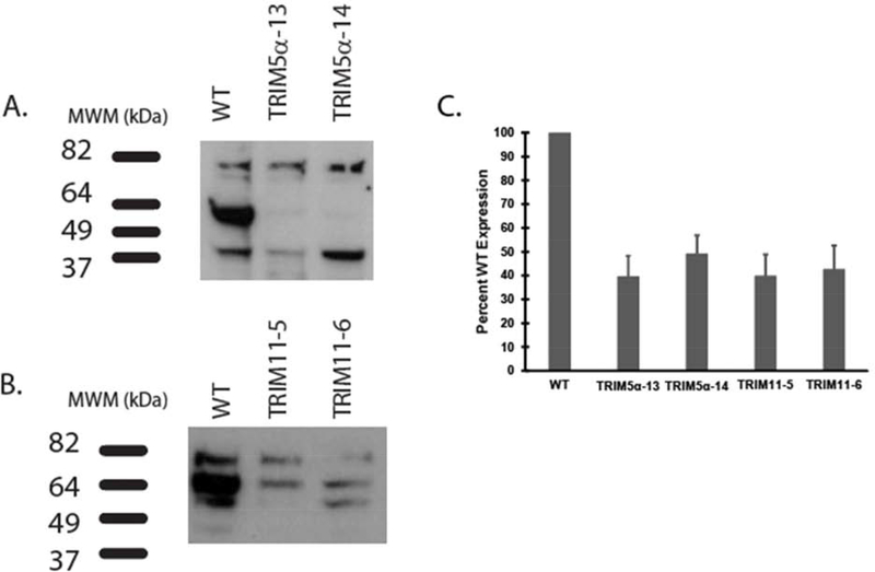
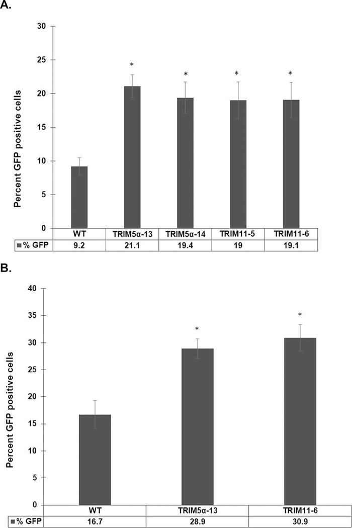
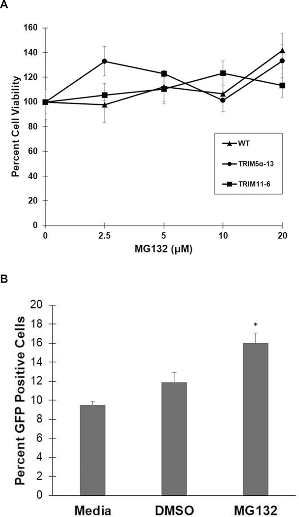
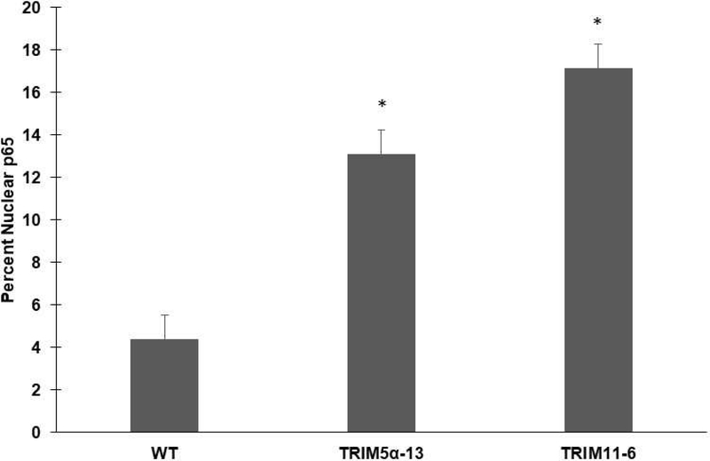
The goal of this study was to determine the expression and distribution of the host restriction factors (RFs) TRIM5α and TRIM11 in non-human primate (NHP) neural retina tissue and the human Muller cell line MIO-M1. In addition, experiments were performed to determine the effect of TRIM5α and TRIM11 knockdown on FIVGFP transduction of MIO-M1 cells with the goal of devising strategies to increase the efficiency of lentiviral (LV) gene delivery. Immunofluorescence (IF) studies indicated that TRIM5α and TRIM11 were localized predominantly in nuclei within the outer nuclear layer (ONL) and inner nuclear layer (INL) of NHP retina tissue. Double label IF indicated that TRIM5α and TRIM11 were localized to some of the retinal Muller cell nuclei. MIO-M1 cells expressed TRIM5α predominantly in the nucleus and TRIM11 primarily in the cytosol. FIVGFP transduction efficiency was significantly increased, at 4 and 7 days post transduction, in TRIM5α and TRIM11 knockdown clones (KD) compared to WT MIO-M1 cells. In addition, pretreatment with the proteasome inhibitor MG132 increased the transduction efficiency of FIVGFP in WT MIO-M1 cells. The nuclear translocation of NF-κB (p65), at 72 h post FIVGFP transduction, was enhanced in TRIM5α and TRIM11 KD clones. The expression of TRIM5α and TRIM11 in macaque neural retina tissue and MIO-M1 cells indicate the presence of these RFs in NHP retina and human Muller cells. Our data indicate that even partial knockdown of TRIM5α or TRIM11, or a short proteasome inhibitor pretreatment, can increase the transduction efficiency of a LV vector.

Keywords: Gene therapy; Muller cells; Proteasome inhibitor; Retina; TRIM11; TRIM5α; Viral vectors.


Figures

Figure 1:
Figure 1:. Expression of TRIM5α and TRIM11 proteins in NHP retina tissue.
Immunoblotting of macaque retina tissue lysates was performed with A) rabbit anti-TRIM5α or B) rabbit anti-TRIM11 primary antibodies. Goat anti-rabbit HRP-conjugated secondary antibodies were applied prior to ECL.
Figure 2:
Figure 2:. Expression patterns of TRIM5α and TRIM11 in macaque neural retina tissue:
A) Arrows indicate TRIM5α staining. B and E) Arrows indicate vimentin staining of Muller cells. C) Merged image with arrows indicating localization of TRIM5α staining to Muller cell nuclei. D) Arrows indicate TRIM11 staining. F) Merged image indicating localization of TRIM11 staining to Muller cell nuclei. PR= photoreceptor layer, ONL= outer nuclear layer, INL= inner nuclear layer, GCL= ganglion cell layer. Scale bar= 20μm.
Figure 3:
Figure 3:. Expression patterns of TRIM5α and TRIM11 in MIO-M1 cells.
MIO-M1 cells were fixed, permeabilized, and stained with A) no primary antibody, B) a nonspecific rabbit anti-FLAG Tag antibody, C) rabbit anti-TRIM5α, or D) rabbit anti-TRIM11 followed by a goat anti-rabbit Alexa Fluor 488 secondary antibody. All polyclonal antibodies were diluted to a final concentration of 10 μg/ml.
Figure 4:
Figure 4:. Immunoblots of TRIM5α and TRIM11 knockdown clones.
Equal micrograms of MIO-M1 WT and TRIM5α, or MIO-M1 WT and TRIM11 knockdown cell lysates, were electrophoresed and blotted with: A) mouse anti-TRIM5 or B) rabbit anti-TRIM11 antibodies. HRP-conjugated secondary antibodies were applied prior to ECL. C) ImageJ analysis was performed on three separate blots for each antibody and the mean percent WT expression was graphed with error bars representing standard error of the mean (SEM).
Figure 5:
Figure 5:. TRIM5α or TRIM11 knockdown increased FIVGFP transduction efficiency in a human Muller cell line.
WT MIO-M1, TRIM5α clones 13 and 14, and TRIM11 clones 5 and 6 were challenged with FIVGFP. Four days (A) or seven days (B) post transduction, cells were fixed and stained with an anti-TurboGFP antibody followed by goat anti-rabbit Alexa Fluor 488. Cells were stained with Hoechst to visualize nuclei. The percent GFP positive cells were determined in five fields for each cell type. Error bars represent SEM. p values were determined by two-sample t-test. Sample size (N) = 5. (*): p<0.02.
Figure 6:
Figure 6:. Proteasome inhibitor pretreatment increased FIVGFP transduction efficiency in MIO-M1 cells.
A) WT MIO-M1, TRIM5α−13 KD cells, and TRIM11–6 KD cells were incubated with increasing concentrations of MG132 for 1 hour at 37°C, followed by MTS assay. The mean percent viability was calculated and graphed with error bars representing the SEM. B) WT MIO-M1 cells were pretreated with media, 0.05% DMSO, or 20 μM MG132/0.05% DMSO for 1 hour at 37°C prior to FIVGFP transduction (MOI 20). Six days post transduction, the cells were fixed and stained with an anti-TurboGFP antibody followed by goat anti-rabbit AlexaFluor 488. Cells were stained with Hoechst to visualize nuclei. The percent GFP positive cells were determined in five fields for each condition and the mean percent was graphed with error bars representing the SEM. p values were determined by two-sample t-test. Sample size (N)= 5. ( *): p=0.002.
Figure 7:
Figure 7:. NF-κB (p65) activation following FIVGFP transduction
Duplicate wells of WT, TRIM5α−13 KD, and TRIM11–6 KD MIO-M1 cells were challenged with FIVGFP for 72 hours. Cells were fixed, permeabilized, and stained with an anti-NF-κB (p65) antibody followed by donkey anti-goat Alexa Fluor 594. Cells were stained with Hoechst to visualize nuclei. The total number of cells and p65 positive nuclei were determined in five random fields per well and data was graphed as mean percent nuclear p65. Error bars represent SEM. p values were determined by two-sample t-test. (*): p < 0.001. Sample size (N)=10

Similar articles

Cited by

References

    1. Aktas Z, Rao H, Slauson SR, Gabelt BT, Larsen IV, Sheridan RTC, Herrnberger L, Tamm ER, Kaufman PL, Brandt CR: Proteasome inhibition increases the efficiency of lentiviral vector-mediated transduction of trabecular meshwork. Invest. Ophthalmol. Vis. Sci, 59 (2018), pp. 298–310, 10.1167/iovs.17-22074. - DOI - PMC - PubMed
    1. Annoni A, Gregori S, Naldini L, Cantore A: Modulation of immune responses in lentiviral vector-mediated gene transfer. Cell. Immunol, 342 (2019), pp. 103802, 10.1016/j.cellimm.2018.04.012. - DOI - PMC - PubMed
    1. Balaggan KS, Ali RR: Ocular gene delivery using lentiviral vectors. Gene Ther, 19 (2012), pp. 145–153, 10.1038/gt.2011.153. - DOI - PubMed
    1. Balaggan KS, Binley K, Esapa M, Iqball S, Askham Z, Kan O, Tschernutter M, Bainbridge JW, Naylor S, Ali RR: Stable and efficient intraocular gene transfer using pseudotyped EIAV lentiviral vectors. J. Gene Med, 8 (2006), pp. 275–285, 10.1002/jgm.845. - DOI - PubMed
    1. Bennett J: Taking stock of retinal gene therapy: looking back and moving forward. Mol. Ther, 25 (2017), pp. 1076–1094, 10.1016/j.ymthe.2017.03.008. - DOI - PMC - PubMed

Publication types

MeSH terms