Review
. 2021 Jan 11;11(1):101.
doi: 10.3390/diagnostics11010101.

From Nutcracker Phenomenon to Nutcracker Syndrome: A Pictorial Review

Affiliations
Review

From Nutcracker Phenomenon to Nutcracker Syndrome: A Pictorial Review

Antonio Granata et al. Diagnostics (Basel). .

Abstract

Left renal vein (LRV) entrapment, also known as nutcracker phenomenon if it is asymptomatic, is characterized by abnormality of outflow from the LRV into the inferior vena cava (IVC) due to extrinsic LRV compression, often accompanied by demonstrable lateral (hilar) dilatation and medial (mesoaortic) stenosis. Nutcracker syndrome, on the other hand, includes a well-defined set of symptoms, and the severity of these clinical manifestations is related to the severity of anatomic and hemodynamic findings. With the aim of providing practical guidance for nephrologists and radiologists, we performed a review of the literature through the PubMed database, and we commented on the definition, the main clinical features, and imaging pattern of this syndrome; we also researched the main therapeutic approaches validated in the literature. Finally, from the electronic database of our institute, we have selected some characteristic cases and we have commented on the imaging pattern of this disease.

Keywords: CT; ECD; nutcracker phenomenon; nutcracker syndrome; renal; renovascular hypertension.


Conflict of interest statement

The authors declare no conflict of interest.

Figures

Figure 1
Figure 1
3D volume rendering (VR) contrast enhanced CT scan (portal venous phase) shows the relationships between the aorta, the superior mesenteric artery, and the left renal vein in a patient with nutcracker syndrome; in this acquisition, an initial enhancement of the left gonadal vein is also appreciable, which appears dilated (varicocele).
Figure 2
Figure 2
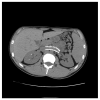
A patient with nutcracker syndrome: contrast enhanced CT scan during arterial phase, with sagittal reconstruction; in this image, it is possible to appreciate the entrapment of the left renal vein (white arrow) between the superior mesenteric artery and the aorta.
Figure 3
Figure 3
Maximum intensity projection (MIP) = 25 mm and multi-planar reformation (MPR), (a) sagittal, (b) axial, and (c) coronal contrast enhanced CT portal venous phase images of a young woman affected by nutcracker syndrome with pelvic congestion syndrome, acquired in portal venous phase; in (a) and in (c), pathological serpiginous dilatation of the left ovarian vein is shown.
Figure 4
Figure 4
Another patient with nutcracker syndrome and pelvic congestion syndrome; angiographic acquisition shows left pelvic varicocele, characterized by appreciable dilatation of the left ovarian vein.
Figure 5
Figure 5
Patient affected by nutcracker syndrome. The sagittal reconstruction demonstrates an aorto-mesenteric angle of about 33°, lower than the norm (aorto-mesenteric angle normally ranges from 38° to 56°).
Figure 6
Figure 6
Axial view of contrast enhancement CT scan demonstrating the beak sign (white arrow), caused by the compression of the left renal vein from the aorta and superior mesenteric artery.
Figure 7
Figure 7
Another nutcracker syndrome patient; in this axial image, it is possible to appreciate a beck angle higher than 32°, suggestive of entrapment of the left renal vein.
Figure 8
Figure 8
A left renal vein diameter ratio higher than 4.9 is specific for nutcracker syndrome; it is necessary to measure the axial diameter of the renal vein at the renal hilum and in correspondence with the stenosis between the superior mesenteric artery and the aorta.
Figure 9
Figure 9
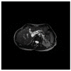
RMI, axial FIESTA sequence, shows the compression of the left renal vein between the aorta and superior mesenteric artery in a patient affected by nutcracker syndrome with flank pain and pelvic congestion syndrome. In this image, the beck sign (white arrow) is highlighted; SMA: superior mesenteric artery, A: aorta, LRV: left renal vein.
Figure 10
Figure 10
A patient suffering from flank pain, varicocele, and hematuria evaluated at the ultrasound (US) laboratory of the Nephrology and Dialysis Unit. The axial grayscale sonogram demonstrates the anatomical relationships between the left renal vein, aorta, and superior mesenteric artery, with stenosis at the aorto-mesenteric angle (beck sign) and dilation of the hilar section of the left renal vein with a ratio of nutcracker phenomenon. A: aorta, SMA: superior mesenteric artery, LRV: left renal vein, IVC: inferior vena cava, VB: vertebral body, white asterisk: beck sign.
Figure 11
Figure 11
A rare case of posterior nutcracker syndrome evaluated with US; in this axial image, it is possible to recognize the left renal vein that runs between the abdominal aorta and the vertebral column. A: aorta, LRV: left renal vein, IVC: inferior vena cava, VB: vertebral body.
Figure 12
Figure 12
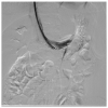
Patient affected by nutcracker syndrome (with nutcracker phenomenon, flank pain, hematuria, and left varicocele) treated with endovascular procedures; diagnostic phlebography documented a significant stenosis of the left renal vein at the aorto-mesenteric clap; at the stenosis, a stent (visible in the image) dilated with a balloon catheter was released.
Figure 13
Figure 13
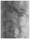
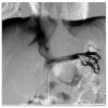
Same patient as the previous figure, re-evaluated a year later for a new episode of hematuria; diagnostic phlebography documents that the stent is in the normal position and is normally patent; collateral circulation or varicose ectasias are not recognizable.
Figure 14
Figure 14
Patient who previously underwent endovascular treatment with symptom resolution, subjected to imaging investigations for other reasons. Non-contrast axial CT passing through aorto-mesenteric clamp demonstrated the correct positioning of a stent in the left renal vein with resolution of the stenosis.

References

    1. Lamba R., Tanner D., Sekhon S., McGahan J., Corwin M., Lall C. Multidetector CT of vascular compression syndromes in the abdomen and pelvis. Radiographics. 2014;34:93–115. doi: 10.1148/rg.341125010. - DOI - PubMed
    1. Eliahou R., Sosna J., Bloom A.I. Between a Rock and a Hard Place: Clinical and Imaging Features of Vascular Compression Syndromes. Radiographics. 2012;32:E33–E49. doi: 10.1148/rg.321115011. - DOI - PubMed
    1. Kurklinsky A.K., Rooke T.W. Nutcracker Phenomenon and Nutcracker Syndrome. Mayo Clin. Proc. 2010;85:552–559. doi: 10.4065/mcp.2009.0586. - DOI - PMC - PubMed
    1. Shin J.I., Lee J.S. Nutcracker phenomenon or nutcracker syndrome? Nephrol. Dial. Transplant. 2005;20:2009. doi: 10.1093/ndt/gfi078. - DOI - PubMed
    1. Cope C., Isard H.J. Left renal vein entrapment: A new diagnostic finding in retroperitoneal disease. Radiology. 1969;92:867–872. doi: 10.1148/92.4.867. - DOI - PubMed

LinkOut - more resources