

The Neurosurgeon's Armamentarium for Gliomas: An Update on Intraoperative Technologies to Improve Extent of Resection
- PMID: 33440712
- PMCID: PMC7826675
- DOI: 10.3390/jcm10020236
The Neurosurgeon's Armamentarium for Gliomas: An Update on Intraoperative Technologies to Improve Extent of Resection
Abstract
Maximal safe resection is the standard of care in the neurosurgical treatment of high-grade gliomas. To aid surgeons in the operating room, adjuvant techniques and technologies centered around improving intraoperative visualization of tumor tissue have been developed. In this review, we will discuss the most advanced technologies, specifically fluorescence-guided surgery, intraoperative imaging, neuromonitoring modalities, and microscopic imaging techniques. The goal of these technologies is to improve detection of tumor tissue beyond what conventional microsurgery has permitted. We describe the various advances, the current state of the literature that have tested the utility of the different adjuvants in clinical practice, and future directions for improving intraoperative technologies.
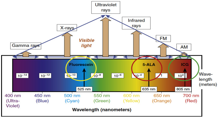
Keywords: 5-ALA; extent of resection; fluorescein; fluorescence-guided surgery; glioblastoma; glioma surgery; iMRI; intraoperative imaging; neuromonitoring; ultrasound.
Conflict of interest statement
C.G.H. is a consultant for NX Development Corporation (NXDC) and Synaptive Medical. NXDC, a privately held company, markets Gleolan (5-ALA, aminolevulinic acid hydrochloride). Gleolan is an optical imaging agent approved for the visualization of malignant tissue during glioma surgery. Hadjipanayis is a consultant for NXDC and receives royalty payments for the sale of Gleolan. Hadjipanayis receives financial compensation as a consultant and lecturer for Synaptive (manufacturer of the 3D Synaptive MODUS V device). He has also received speaker fees by Carl Zeiss and Leica.
Figures

References
-
- Lacroix M., Abi-Said D., Fourney D.R., Gokaslan Z.L., Shi W., DeMonte F., Lang F.F., McCutcheon I.E., Hassenbusch S.J., Holland E., et al. A multivariate analysis of 416 patients with glioblastoma multiforme: Prognosis, extent of resection, and survival. J. Neurosurg. 2001;95:190–198. doi: 10.3171/jns.2001.95.2.0190. - DOI - PubMed
-
- McGirt M.J., Chaichana K.L., Gathinji M., Attenello F.J., Than K., Olivi A., Weingart J.D., Brem H., Quiñones-Hinojosa A.R. Independent association of extent of resection with survival in patients with malignant brain astrocytoma. J. Neurosurg. 2009;110:156–162. doi: 10.3171/2008.4.17536. - DOI - PubMed
-
- Hadjipanayis C.G., Stummer W. Fluorescence-Guided Neurosurgery: Neuro-Oncology and Cerebrovascular Applications. Thieme; New York, NY, USA: 2019.
Publication types
LinkOut - more resources
Full Text Sources
Other Literature Sources
