. 2021 Jan 11;13(1):202.
doi: 10.3390/nu13010202.

Bifidobacterium breve CNCM I-4035, Lactobacillus paracasei CNCM I-4034 and Lactobacillus rhamnosus CNCM I-4036 Modulate Macrophage Gene Expression and Ameliorate Damage Markers in the Liver of Zucker-Lepr fa/fa Rats

Affiliations

Bifidobacterium breve CNCM I-4035, Lactobacillus paracasei CNCM I-4034 and Lactobacillus rhamnosus CNCM I-4036 Modulate Macrophage Gene Expression and Ameliorate Damage Markers in the Liver of Zucker-Lepr fa/fa Rats

Luis Fontana et al. Nutrients. .

Abstract

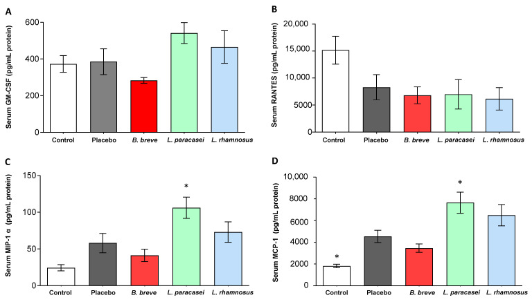
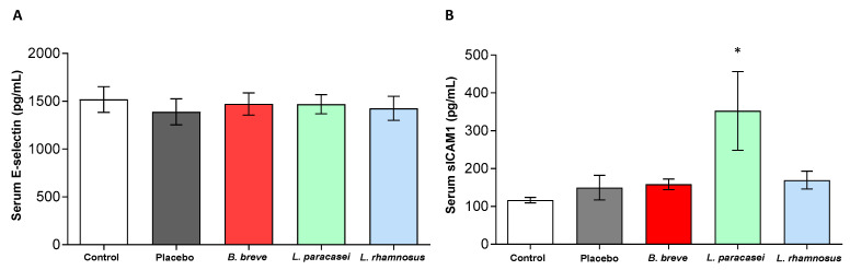
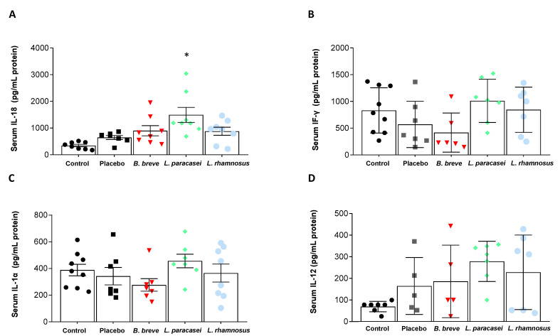
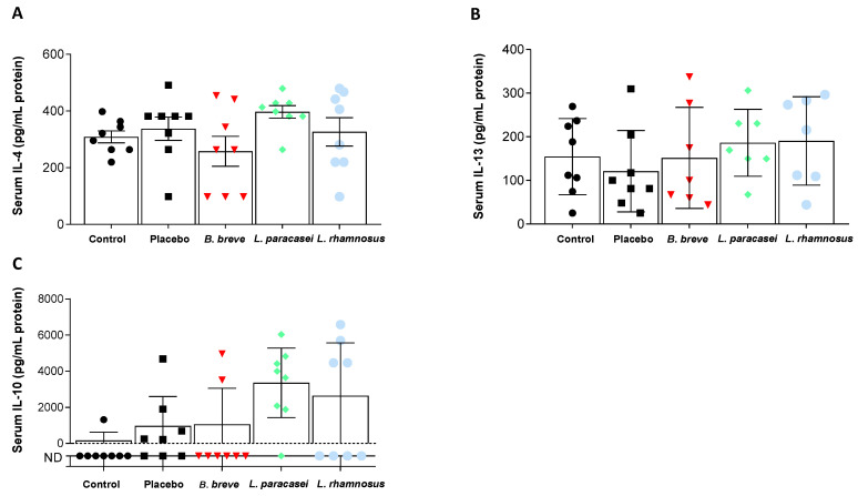
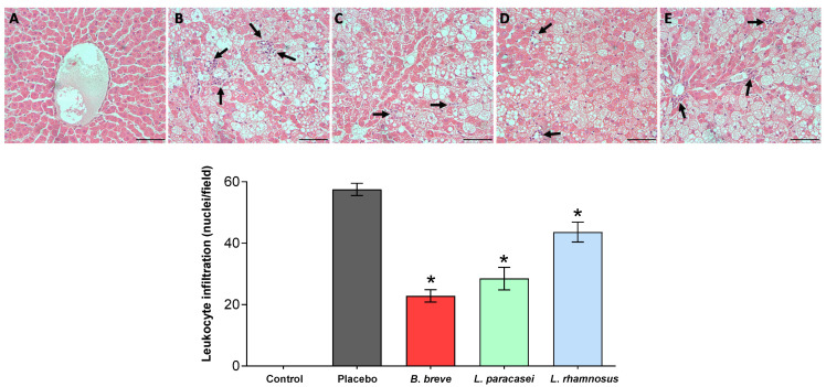
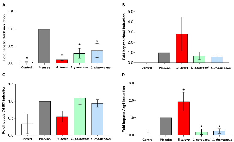
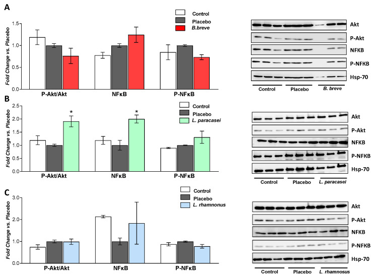
Non-alcoholic fatty liver disease (NAFLD) has reached pandemic proportions worldwide. We have previously reported that the probiotic strains Bifidobacterium breve CNCM I-4035, Lactobacillus paracasei CNCM I-4034 and Lactobacillus rhamnosus CNCM I-4036 exert anti-inflammatory effects in the intestine of Zucker-Lepr fa/fa rats. In this work, we focused on their hepatic effects. M1 macrophages are related to inflammation and NAFLD pathogenesis, whereas M2 macrophages release anti-inflammatory mediators. We evaluated the effects of these 3 strains on macrophage polarization, inflammation and liver damage of Zucker-Lepr fa/fa rats. The animals received either a placebo or 1010 CFU of probiotics orally for 30 days. Nos2 and Cd86 mRNA levels were determined as markers of M1 macrophages, and Cd163 and Arg1 as M2 markers, respectively, by qRT-PCR. Liver damage was determined by lipid peroxidation, leukocyte infiltration and myeloperoxidase activity. We evaluated a panoply of circulating chemokines, the hepatic ratio P-Akt/Akt, NF-kB and P-NF-kB protein levels. All 3 probiotic strains modulated macrophage polarization in liver and circulating levels of inflammation-related mediators. L. paracasei CNCM I-4034 increased the ratio P-Akt/Akt and NF-kB protein levels. B. breve CNCM I-4035, L. paracasei CNCM I-4034 and L. rhamnosus CNCM I-4036 decreased both pro-inflammatory macrophage gene expression and leukocyte infiltration in the liver.

Keywords: NAFLD; inflammation; liver damage; macrophages; polarization; probiotics.


Conflict of interest statement

The authors declare no conflict of interest.

Figures

Figure 1
Figure 1
Serum concentration of (A) GM-CSF, (B) RANTES, (C) MIP-1 α, and (D) MCP-1 of rats fed either placebo, B. breve CNCM I-4035, L. paracasei CNCM I-4034 or L. rhamnosus CNCM I-4036 for 30 days. Values are the means ± SEM. n = 8 per group. * p < 0.05 vs. placebo. B, Bifidobacterium; GM-CSF, granulocyte-macrophage colony-stimulating factor; L, Lactobacillus; MCP-1, monocyte chemoattractant protein-1; MIP-1 α, macrophage inflammatory protein 1-alpha;RANTES, Regulated upon Activation, Normal T Cell Expressed and Presumably Secreted.
Figure 2
Figure 2
Serum concentration of (A) E-Selectin and (B) sICAM1 of rats fed either placebo, B. breve CNCM I-4035, L. paracasei CNCM I-4034 or L. rhamnosus CNCM I-4036 for 30 days. Values are the means ± SEM. n = 8 per group. * p < 0.05 vs. placebo. B, Bifidobacterium; L, Lactobacillus; sICAM1: soluble intercellular adhesion molecule-1.
Figure 3
Figure 3
Serum concentration of pro-inflammatory cytokines. (A) IL-18, (B) INF-γ, (C) IL-1α, and (D) IL-12 of rats fed either placebo, B. breve CNCM I-4035, L. paracasei CNCM I-4034 or L. rhamnosus CNCM I-4036 for 30 days. Values are the means ± SEM. n = 8 per group. * p < 0.05 vs. placebo. B, Bifidobacterium; INF-γ, interferon-gamma; IL, interleukin; L. Lactobacillus.
Figure 4
Figure 4
Serum concentration of anti-inflammatory cytokines. (A) IL-4, (B) IL-13, and (C) IL-10 of rats fed either placebo, B. breve CNCM I-4035, L. paracasei CNCM I-4034 or L. rhamnosus CNCM I-4036 for 30 days. Values are the means ± SEM. n = 8 per group. B, Bifidobacterium; IL, interleukin; L. Lactobacillus.
Figure 5
Figure 5
Inflammatory cell infiltration. Upper panels: Representative micrographs of 5-µm-thick liver sections stained with hematoxylin and eosin (H&E) of (A) control group, (B) placebo group, (C) B. breve group, (D) L. paracasei group, and (E) L. rhamnosus group. Bars = 20 µm. Lower panel: Quantification corresponding to the stained sections. Values are the means ± SEM, n = 3–4 per group. * p < 0.05 vs. placebo. B, Bifidobacterium; L, Lactobacillus.
Figure 6
Figure 6
Hepatic levels of (A) MPO, (C) MDA and (D) LPS, and serum levels of MPO (B) of rats fed either with placebo, B. breve CNCM I-4035, L. paracasei CNCM I-4034 or L. rhamnosus CNCM I-4036 for 30 days. Values are the means SEM, n = 3–5 per group. * p < 0.05 vs. placebo. B, Bifidobacterium; L, Lactobacillus; LPS, lipopolysaccharide; MDA, malondialdehyde; MPO, myeloperoxidase.
Figure 7
Figure 7
Expression of macrophage genes in liver. M1 macrophage markers: (A) Cd86 and (B) Nos2. M2 macrophage markers: (C) Cd163 and (D) Arg1 of rats fed either placebo or B. breve CNCM I-4035, L. paracasei CNCM I-4034 or L. rhamnosus CNCM I-4036 for 30 days. Relative mRNA levels were normalized to the trancript levels of the housekeeping gene Hprt1. Data were calculated as fold change compared with the placebo group. Values are the means ± SEM, n = 3–5 per group. * p < 0.05 vs. placebo. Arg1, Arginase-1; B, Bifidobacterium; Cd, cluster of differentiation; L, Lactobacillus; Nos2, Nitric oxide synthase-2.
Figure 8
Figure 8
Western blot analysis of P-Akt/Akt ratio, NF-kB and P-NF-kB proteins of rats fed either placebo, (A) B. breve, (B) L. paracasei or (C) L. rhamnosus for 30 days. Hsp-70 was used as a loading control. Graphs on the left included 3 rats per group, and the right panels show representative blots. Values are the means ± SEM. * p < 0.05 vs. placebo. Akt, protein kinase B; B, Bifidobacterium; Hsp70, 70 KD heat shock protein; L, Lactobacillus. NF-κB, nuclear factor kappa-light-chain-enhancer of activated B cells; P-Akt, phosphorylated protein kinase B; P-NF-kB, phosphorylated nuclear factor kappa-light-chain-enhancer of activated B cells.

Similar articles

Cited by

References

    1. Di Sessa A., Cirillo G., Guarino S., Marzuillo P., Del Giudice E.M. Pediatric non-alcoholic fatty liver disease: Current perspectives on diagnosis and management. Pediatr. Health Med. Ther. 2019;10:89–97. doi: 10.2147/PHMT.S188989. - DOI - PMC - PubMed
    1. Álvarez-Mercado A.I., Gulfo J., Romero Gómez M., Jiménez-Castro M.B., Gracia-Sancho J., Peralta C. Use of steatotic grafts in liver transplantation: Current status. Liver Transpl. 2019;25:771–786. doi: 10.1002/lt.25430. - DOI - PubMed
    1. Benedict M., Zhang X. Non-alcoholic fatty liver disease: An expanded review. World J. Hepatol. 2017;9:715–732. doi: 10.4254/wjh.v9.i16.715. - DOI - PMC - PubMed
    1. Meli R., Raso G.M., Calignano A. Role of Innate Immune Response in Non-Alcoholic Fatty Liver Disease: Metabolic Complications and Therapeutic Tools. Front. Immunol. 2014;5:177. doi: 10.3389/fimmu.2014.00177. - DOI - PMC - PubMed
    1. Fraternale A., Brundu S., Magnani M. Polarization and repolarization of macrophages. J. Clin. Cell Immunol. 2015;6:2.