

Ex Vivo Live Full-Thickness Porcine Skin Model as a Versatile In Vitro Testing Method for Skin Barrier Research
- PMID: 33440780
- PMCID: PMC7827261
- DOI: 10.3390/ijms22020657
Ex Vivo Live Full-Thickness Porcine Skin Model as a Versatile In Vitro Testing Method for Skin Barrier Research
Abstract
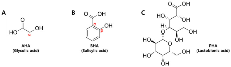
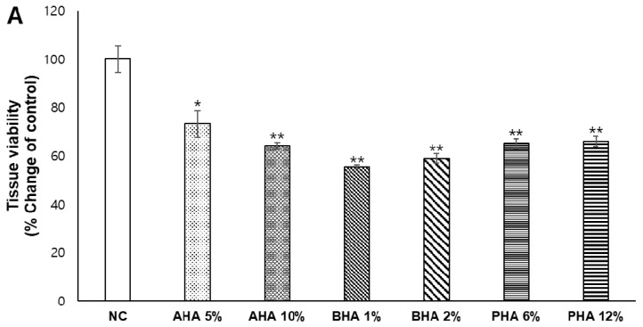
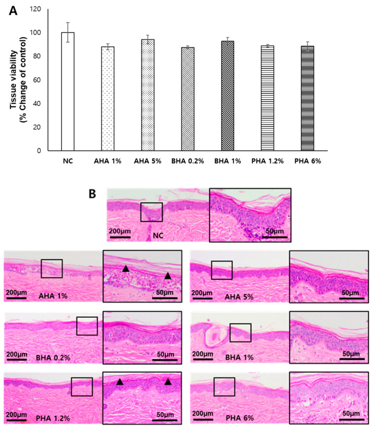
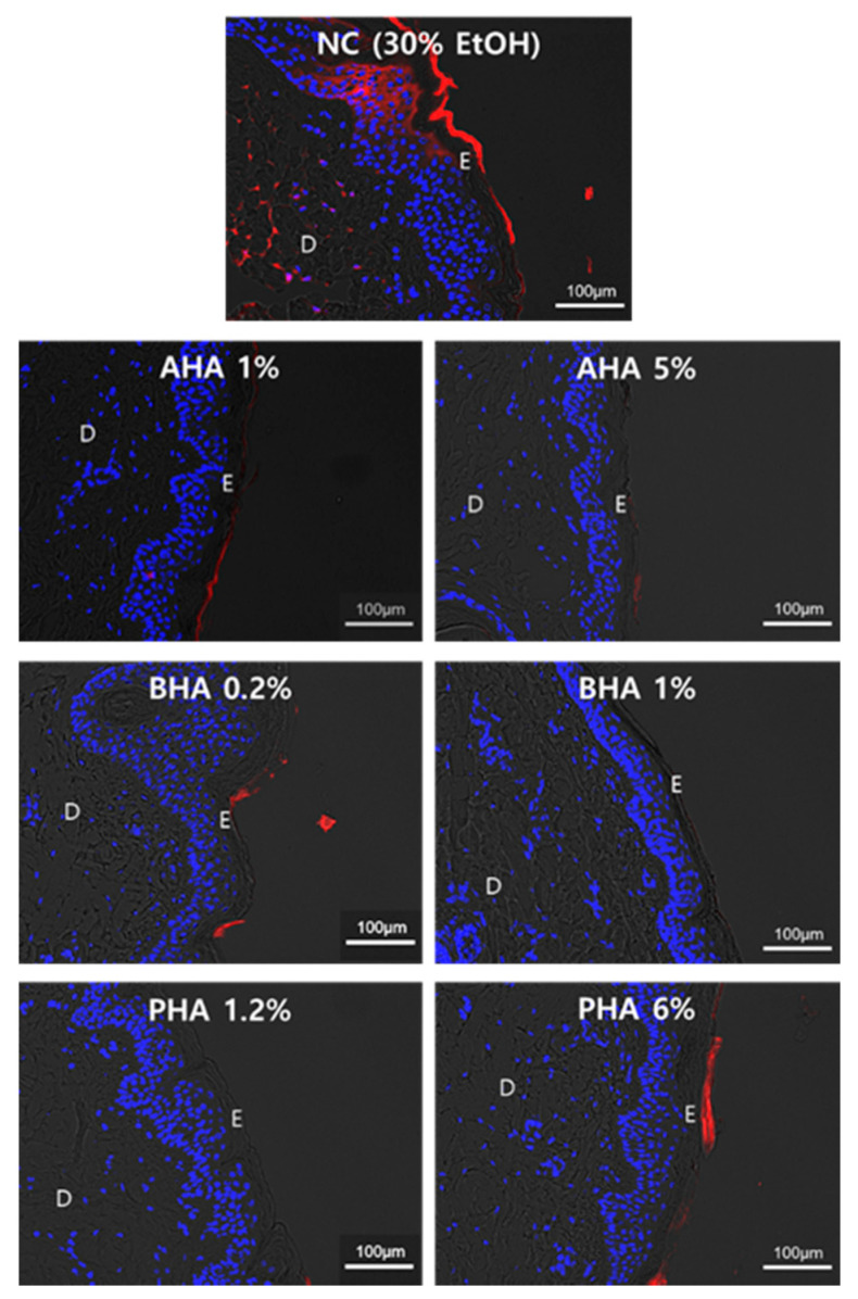
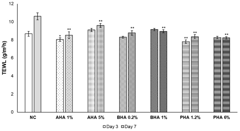
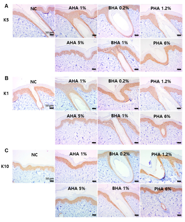
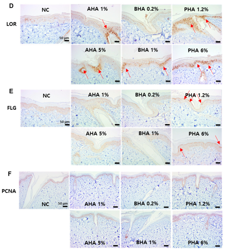
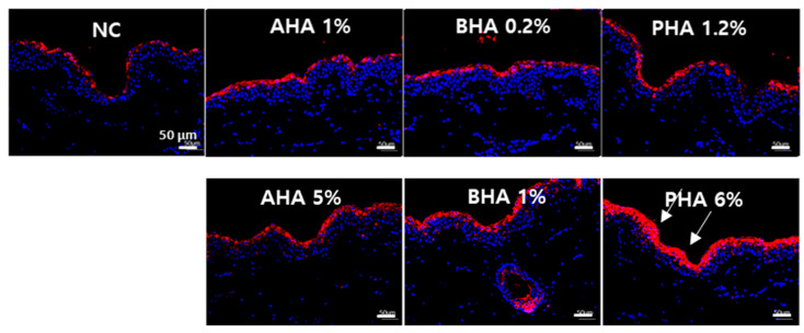
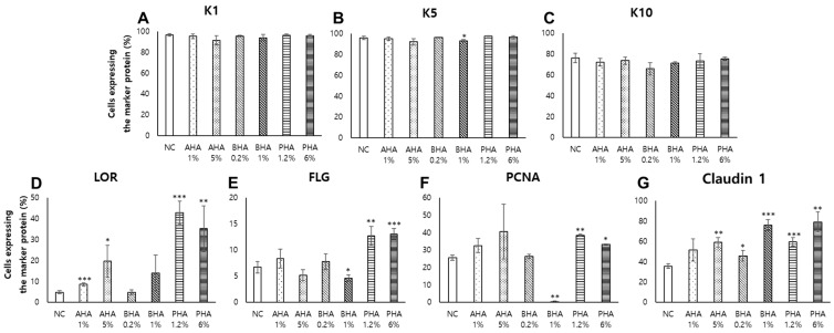
Since the European Union (EU) announced their animal testing ban in 2013, all animal experiments related to cosmetics have been prohibited, creating a demand for alternatives to animal experiments for skin studies. Here, we investigated whether an ex vivo live porcine skin model can be employed to study the safety and skin barrier-improving effects of hydroxyacids widely used in cosmetics for keratolytic peels. Glycolic acid (1-10%), salicylic acid (0.2-2%), and lactobionic acid (1.2-12%) were used as representative substances for α-hydroxyacid (AHA), β-hydroxyacid (BHA), and polyhydroxyacid (PHA), respectively. When hydroxyacids were applied at high concentrations on the porcine skin every other day for 6 days, tissue viability was reduced to 50-80%, suggesting that the toxicity of cosmetic ingredients can be evaluated with this model. Based on tissue viability, the treatment scheme was changed to a single exposure for 20 min. The protective effects of a single exposure of hydroxyacids on skin barrier function were evaluated by examining rhodamine permeability and epidermal structural components of barrier function using immunohistochemistry (IHC) and immunofluorescence (IF) staining. Lactobionic acid (PHAs) improved skin barrier function most compared to other AHAs and BHAs. Most importantly, trans-epidermal water loss (TEWL), an important functional marker of skin barrier function, could be measured with this model, which confirmed the significant skin barrier-protective effects of PHAs. Collectively, we demonstrated that the ex vivo live full-thickness porcine skin model can be an excellent alternative to animal experiments for skin studies on the safety and efficacy of cosmetic ingredients.
Keywords: ex vivo skin model; hydroxyacids; skin barrier; skin permeability; stratum corneum.
Conflict of interest statement
The authors declare no conflict of interest.
Figures
Similar articles
-
Alpha hydroxyacids modulate stratum corneum barrier function.Br J Dermatol. 1997 Dec;137(6):934-8. Br J Dermatol. 1997. PMID: 9470910 Clinical Trial.
-
Photocarcinogenesis study of glycolic acid and salicylic acid (CAS Nos. 79-14-1 and 69-72-7) in SKH-1 mice (simulated solar light and topical application study).Natl Toxicol Program Tech Rep Ser. 2007 Sep;(524):1-242. Natl Toxicol Program Tech Rep Ser. 2007. PMID: 21921960
-
The effects of topical alpha-hydroxyacids on the normal skin barrier of hairless mice.Br J Dermatol. 2001 Feb;144(2):267-73. doi: 10.1046/j.1365-2133.2001.04011.x. Br J Dermatol. 2001. PMID: 11251557
-
The use of polyhydroxy acids (PHAs) in photoaged skin.Cutis. 2004 Feb;73(2 Suppl):3-13. Cutis. 2004. PMID: 15002656 Review.
-
Clinical and cosmeceutical uses of hydroxyacids.Clin Dermatol. 2009 Sep-Oct;27(5):495-501. doi: 10.1016/j.clindermatol.2009.06.023. Clin Dermatol. 2009. PMID: 19695482 Review.
Cited by
-
Assessing Anti-Acne Potentials Via In vitro, Ex vivo, and In vivo Models: A Comprehensive Approach.Curr Drug Targets. 2025;26(7):435-453. doi: 10.2174/0113894501335548250123072644. Curr Drug Targets. 2025. PMID: 39886785 Review.
-
Biopolymer Nano-Network for Antimicrobial Peptide Protection and Local Delivery.Adv Healthc Mater. 2022 Apr;11(7):e2101426. doi: 10.1002/adhm.202101426. Epub 2022 Jan 12. Adv Healthc Mater. 2022. PMID: 34936732 Free PMC article.
-
Methodologies to Evaluate the Hair Follicle-Targeted Drug Delivery Provided by Nanoparticles.Pharmaceutics. 2023 Jul 21;15(7):2002. doi: 10.3390/pharmaceutics15072002. Pharmaceutics. 2023. PMID: 37514188 Free PMC article. Review.
-
Novel ethosomal gel formulation for enhanced transdermal delivery of curcumin and cyclosporine: a preclinical approach to rheumatoid arthritis management.Drug Deliv. 2025 Dec;32(1):2512620. doi: 10.1080/10717544.2025.2512620. Epub 2025 Jun 3. Drug Deliv. 2025. PMID: 40458935 Free PMC article.
-
Development of an Oral Epithelial Ex Vivo Organ Culture Model for Biocompatibility and Permeability Assessment of Biomaterials.Bioengineering (Basel). 2024 Oct 17;11(10):1035. doi: 10.3390/bioengineering11101035. Bioengineering (Basel). 2024. PMID: 39451410 Free PMC article.
References
-
- Forslind B. A domain mosaic model of the skin barrier. Acta Dermato-Venereol. 1994;74:1–6. - PubMed
-
- Choi J.H., Jin S.W., Lee G.H., Cho S.M., Jeong H.G. Orostachys japonicus ethanol extract inhibits 2,4-dinitrochlorobenzene-induced atopic dermatitis-like skin lesions in NC/Nga mice and TNF-alpha/IFN-gamma-induced TARC expression in HaCaT cells. Toxicol. Res. 2020;36:99–108. doi: 10.1007/s43188-019-00026-0. - DOI - PMC - PubMed
-
- Barel A.O., Paye M., Maibach H.I. Handbook of Cosmetic Science and Technology. CRC Press; Boca Raton, FL, USA: 2014.
-
- Duracher L., Visdal-Johnsen L., Mavon A., Oriflame Cosmetics A.B. Novel Explant Model for Skin Delivery Assessment. Cosm. Toil. 2015;130:30–40.
MeSH terms
Substances
Grants and funding
LinkOut - more resources
Full Text Sources
Other Literature Sources
Miscellaneous
