Review
. 2021 Jan 11;22(2):658.
doi: 10.3390/ijms22020658.

The Involvement of Innate and Adaptive Immunity in the Initiation and Perpetuation of Sjögren's Syndrome

Affiliations
Review

The Involvement of Innate and Adaptive Immunity in the Initiation and Perpetuation of Sjögren's Syndrome

Clara Chivasso et al. Int J Mol Sci. .

Abstract

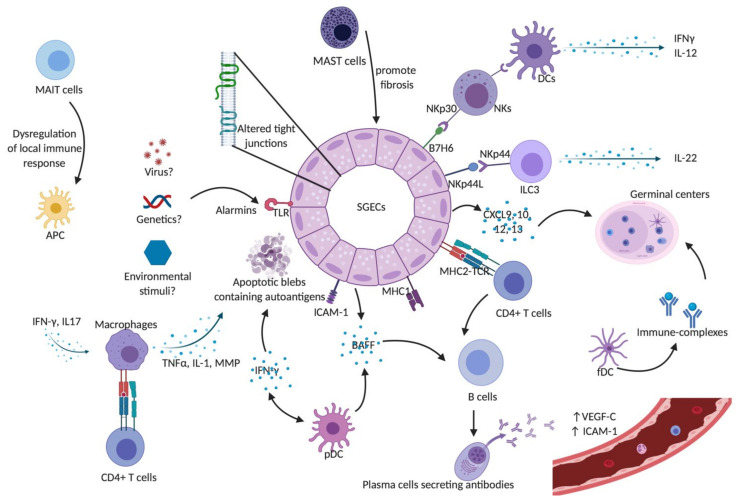
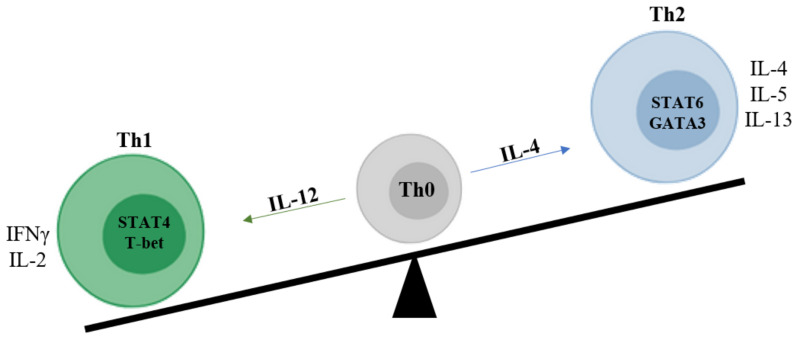
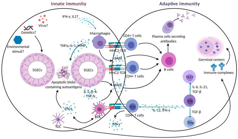
Sjogren's syndrome (SS) is a chronic autoimmune disease characterized by the infiltration of exocrine glands including salivary and lachrymal glands responsible for the classical dry eyes and mouth symptoms (sicca syndrome). The spectrum of disease manifestations stretches beyond the classical sicca syndrome with systemic manifestations including arthritis, interstitial lung involvement, and neurological involvement. The pathophysiology underlying SS is not well deciphered, but several converging lines of evidence have supported the conjuncture of different factors interplaying together to foster the initiation and perpetuation of the disease. The innate and adaptive immune system play a cardinal role in this process. In this review, we discuss the inherent parts played by both the innate and adaptive immune system in the pathogenesis of SS.

Keywords: b cells; epithelial cells; innate immunity; lymphocytes; sjogren’s syndrome; t cells.


Conflict of interest statement

The authors declare no conflict of interest.

Figures

Figure 1
Figure 1
Innate immunity in Sjögren’s syndrome. SGECs constitute the main target of auto-immunity in pSS, described as an autoimmune epithelitis. SGECs exhibit a subverted architecture mainly characterized by altered tight junctions. In genetic susceptible subjects, environmental stimuli such as viruses may trigger salivary gland epithelial cells (SGECs) through TLR activation. Activated SGECs secrete the BAFF that promotes activation and maturation of B cells. SGECs also produce chemokines such as CXCR9, 10, 11, and 12 that attract immune cells and contribute to the formation of germinal centers. Activated SGECs have the ability to act as non-professional antigen-presenting cells by expressing MHC-I, (HLA-ABC) and MHC-II (HLA-DR), adhesion molecules such as ICAM1 allowing them to activate T cells. TLR activation also contributes to SGEC apoptosis, releasing autoantigens that drive autoimmunity in pSS. Activated macrophages produce inflammatory cytokines such as IL-1, TNFα, and MMPs leading to epithelial cell damage. They can also act as antigenic peptide presenting cells through their MHC-II and interact with antigen-specific CD4+ T cells. pDCs lead to the production of type I IFN that acts through autocrine and paracrine circuits feeding a continuous reinforcing inflammatory loop. It also induces the production of BAFF, production contributing to the activation of B cells into plasma cells. fDCs play an essential part in the structure of ectopic germinal centers and retain on their surface immune-complexes, formed by antigen-antibody-complement. Mast cells contribute to the fibrosis and fatty infiltration of salivary glands (SGs). The aberrant phenotype of MAIT cells in pSS patients may lead to the dysregulation of the local immune responses, which would trigger local damage in SGs and auto-immunity. NK cells express the NKp30 receptor that is recognized by DCs and lead to the production of Th1 cytokines such as IFN-γ and IL-12. SGECs express B7-H6, the ligand for NKp30. Taken together, this may explain the hyperactivity of NK cells and the cross-talk with SGECs and DCs that lead to a subsequent activation of innate and adaptive immunity. A subset of ILC3 was found to be a major source of IL-22 in SGECs. Abbreviations: APC: Antigen presenting cells; BAFF: B-cell activating factor; CXCL9: C-X-C motif chemokine type 9; CXCL10: C-X-C motif chemokine type 10; CXCL12: C-X-C motif chemokine type 12; CXCL13: C-X-C motif chemokine type 13; DCs: dendritic cells; fDCs: follicular dendritic cells; ICAM-1: intercellular adhesion molecule 1; IFN-γ: interferon gamma; IL-: interleukin; ILC3: innate Lymphoid Cells type 3 MAIT: Mucosal-associated invariant T cells; MHC-I: major histocompatibility complex class I; MHC-II: major histocompatibility complex class II; MMPs: metalloproteases; NK: natural killer cells; NKp44L: NKp44 ligand; pDCs: plasmacytoid dendritic cells; SGECs: salivary glands epithelial cells; TCR: T cell receptor; TLR: Toll like receptor; TNFα: tumor necrosis factor alpha; VEGF-C: vascular endothelial growth factor C.
Figure 2
Figure 2
Th1-Th2 imbalance. Upon T-cell activation, IFN-γ, and IL-12 induce the expression of T-bet and STAT-4, which is involved in the differentiation of naïve CD4+ T cells into Th1 lymphocytes. Th1 cells predominantly produce pro-inflammatory cytokines such as IFN-γ and IL-2. In contrast, IL-4 induces the GATA-3 transcription factor and the consequent polarization of naïve T cells into Th2. Th2 cells produce anti-inflammatory cytokines such as IL-4, IL-5, and IL-13. Several studies have suggested that pSS is related to abnormal Th1 activation and SGs infiltration. It is supported by the presence of elevated levels of IFN-γ in serum and Th1 cells in blood. Furthermore, T cells expressing a high level of IFN-γ and STAT-4 mRNA have been found in SGs from pSS patients. This Th1/Th2 imbalance, generally observed in various chronic inflammatory disorders, is not easily understood because of a limited number of studies. Abbreviations: IFN-γ:interferon gamma; IL-: interleukin; pSS: primary Sjögren’s syndrome; STAT: signal transducer and activator of transcription; T-bet: T-Box Transcription Factor 21; Th1: type 1 helper cells; Th2: type 2 helper cells.
Figure 3
Figure 3
Innate and adaptive crosstalk. Activated SGECs secrete BAFF that promotes the activation and maturation of B cells into long-lasting memory B cells and plasma B cells producing auto-antibodies. SGECs also produce chemokines IL-1, IL-6, IL18, and TNFα that attract immune cells and contribute to the formation of germinal centers. Activated SGECs have the ability to act as non-professional antigen-presenting cells by expressing MHC-I (HLA-ABC) and MHC-II (HLA-DR) adhesion molecules such as ICAM1, allowing them to activate T cells. TLR activation also contributes to SGEC apoptosis, releasing autoantigens that drive autoimmunity in pSS. Activated macrophages can act as antigenic peptide presenting cells through their MHC-II and interact with antigen-specific CD4+ T cells. pDCs lead to the production of type I IFN that acts through autocrine and paracrine circuits feeding a continuous reinforcing inflammatory loop. It also induces BAFF production, contributing to the activation of B cells into plasma cells. DCs also play an essential part in the structure of ectopic germinal centers and retain on their surface immune-complexes, formed by antigen-antibody-complement.

References

    1. Fox R.I., Kang H.I. Pathogenesis of Sjogren’s syndrome. Rheum. Dis. Clin. N. Am. 1992;18:517–538. - PubMed
    1. Fox R.I., Michelson P. Approaches to the treatment of Sjogren’s syndrome. J. Rheumatol. Suppl. 2000;61:15–21. - PubMed
    1. Basbaum A.I. Opioid regulation of nociceptive and neuropathic pain. Clin. Neuropharmacol. 1992;15 Pt A(Suppl. S1):372A. doi: 10.1097/00002826-199201001-00193. - DOI - PubMed
    1. Sullivan D.A. Sex hormones and Sjogren’s syndrome. J. Rheumatol. Suppl. 1997;50:17–32. - PubMed
    1. Nguyen C.Q., Peck A.B. Unraveling the pathophysiology of Sjogren syndrome-associated dry eye disease. Ocul. Surf. 2009;7:11–27. doi: 10.1016/S1542-0124(12)70289-6. - DOI - PMC - PubMed

LinkOut - more resources