. 2022 Feb 1;107(2):446-456.
doi: 10.3324/haematol.2020.269142.

Successful gene therapy of Diamond-Blackfan anemia in a mouse model and human CD34+ cord blood hematopoietic stem cells using a clinically applicable lentiviral vector

Affiliations

Successful gene therapy of Diamond-Blackfan anemia in a mouse model and human CD34+ cord blood hematopoietic stem cells using a clinically applicable lentiviral vector

Yang Liu et al. Haematologica. .

Abstract

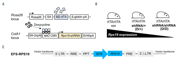
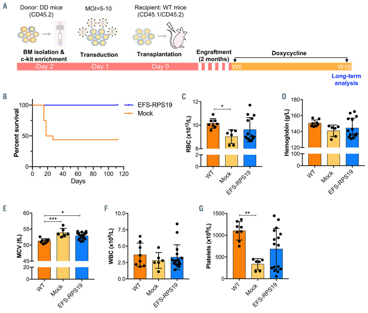
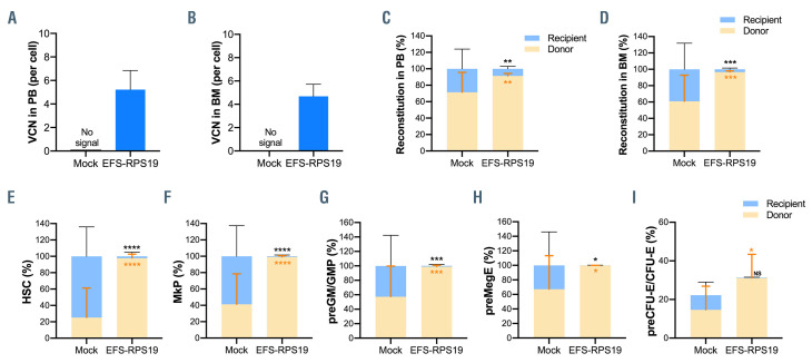
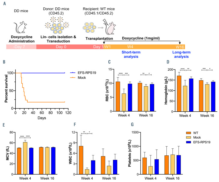
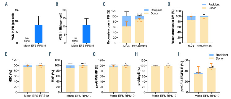
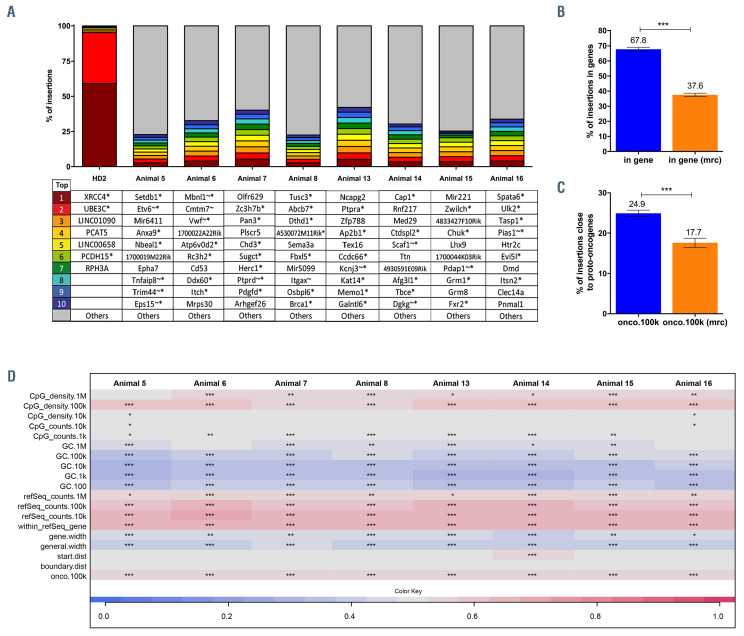
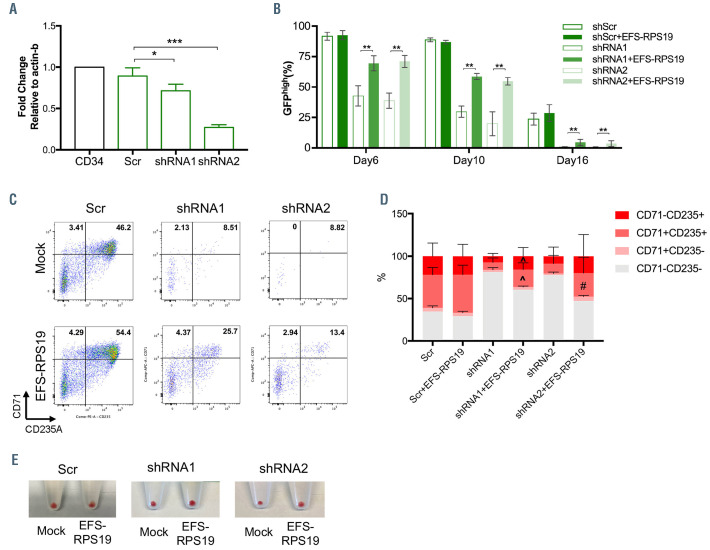
Diamond-Blackfan anemia (DBA) is an inherited bone marrow failure disorder in which pure red blood cell aplasia is associated with physical malformations and a predisposition to cancer. Twentyfive percent of patients with DBA have mutations in a gene encoding ribosomal protein S19 (RPS19). Our previous proof-of-concept studies demonstrated that DBA phenotype could be successfully treated using lentiviral vectors in Rps19-deficient DBA mice. In our present study, we developed a clinically applicable single gene, self-inactivating lentiviral vector, containing the human RPS19 cDNA driven by the human elongation factor 1a short promoter, which can be used for clinical gene therapy development for RPS19-deficient DBA. We examined the efficacy and safety of the vector in a Rps19-deficient DBA mouse model and in human primary RPS19-deficient CD34+ cord blood cells. We observed that transduced Rps19-deficient bone marrow cells could reconstitute mice long-term and rescue the bone marrow failure and severe anemia observed in Rps19-deficient mice, with a low risk of mutagenesis and a highly polyclonal insertion site pattern. More importantly, the vector can also rescue impaired erythroid differentiation in human primary RPS19-deficient CD34+ cord blood hematopoietic stem cells. Collectively, our results demonstrate the efficacy and safety of using a clinically applicable lentiviral vector for the successful treatment of Rps19-deficient DBA in a mouse model and in human primary CD34+ cord blood cells. These findings show that this vector can be used to develop clinical gene therapy for RPS19-deficient DBA patients.


Figures

Figure 1.
Figure 1.
The inducible Rps19-deficient mouse model and structure of the EFS-RPS19 self-inactivating lentiviral vector. (A) Overview of modified loci. Black arrowheads indicate the transcriptional start sites. (B) Breeding strategy to adjust the level of Rps19 downregulation. Homozygous mice (D/D mice) are used in the project. (C) The self-inactivating lentiviral vector harboring a codon-optimized human RPS19 cDNA driven by human elongation factor 1a short (EFS) promoter. LTR: long terminal repeat; pA: polyadenylation signal; PPT: polypurine tract; RRE: Rev response element; SA: splice acceptor.
Figure 2.
Figure 2.
Effective correction of anemia by the EFS-RPS19 vector at 2 weeks after induction of the Diamond-BLackfan phenotype. (A) The scheme of the uninduced genecorrected cell transplantation model and plan for examining short-term therapeutic effects. (B-F) Blood cellularity at 2 weeks after doxycycline induction (n=13-16, error bars represent the standard deviation, *P<0.05, **P<0.01, ***P<0.005 ****P<0.001 by one-way analysis of variance). BM: bone marrow; MOI: multiplicity of infection; WT: wild-type; RBC: red blood cells; MCV: mean corpuscular volume; WBC: white blood cells.
Figure 3.
Figure 3.
Effective long-term correction of the anemia and bone marrow failure in mice treated with the EFS-RPS19 vector. (A) The scheme of the induced genecorrected cell transplantation model and the plan for examining long-term therapeutic effects. (B) Surviral rate analysis. (C-G) Blood cellularity at 16 weeks after doxycycline induction (n=13-16, error bars represent the standard deviation, *P<0.05, **P<0.01, ***P<0.005 by one-way analysis of variance). BM: bone marrow; MOI: multiplicity of infection; WT: wild-type; RBC: red blood cells; MCV: mean corpuscular volume; WBC: white blood cells.
Figure 4.
Figure 4.
Gene-corrected bone marrow cells show a competitive advantage in contributing to long-term hematopoiesis in vivo. (A, B) Vector copy number in peripheral blood (A) and bone marrow (B). (C, D) Donor reconstitution in peripheral blood (C) and bone marrow (D). (E–I) The percentage of transduced cells in hematopoietic stem cells (E), megakaryocyte progenitors (F), pre-granulocyte-macrophage and granulocyte-macrophage progenitors (G), pre-megakaryocyte-erythroid (H), and pre-colony-forming unit erythroid and colony-forming unit erythroid (I) (n=13-16, error bars represent the standard deviation, black asterisks indicate the statistical significance of the comparison of recipient-derived cells between the mock and EFS-RPS19 groups, orange asterisks indicate the statistical significance of the comparison of donor-derived cells between the mock and EFS-RPS19 groups. *P<0.05, **P<0.01, ***P<0.005, ****P<0.001 by one-way analysis of variance). VCN: vector copy number; PB: peripheral blood; BM: bone marrow; HSC: hematopoietic stem cells; MkP: megakaryocyte progenitors; pre-GM/GMP: pre-granulocyte macrophage and granulocyte macrophage progenitors; preMegE: pre-megakaryocyte-erythroid; preCFU-E/CFU-E: pre-colony-forming unit–erythroid (CFU-E)/CFU-E.
Figure 5.
Figure 5.
Amelioration of disease phenotype in Rps19-deficient animals transplanted with gene-corrected cells. (A) Scheme of the gene-corrected Rps19-deficient cell transplantation model and plan for examining short-term and long-term therapeutic effects. (B) Survival rate analysis. (C-G) Blood cellularity at 4 and 16 weeks after doxycycline induction (n=14-16, error bars represent the standard deviation, *P<0.05, **P<0.01, ***P<0.005 ****P<0.001 by one-way analysis of variance). WT: wild-type; RBC: red blood cells; MCV: mean corpuscular volume; WBC: white blood cells.
Figure 6.
Figure 6.
EFS-RPS19 vector-treated Rps19-deficient cells show a competitive advantage in contributing to long-term hematopoiesis in vivo. (A, B) Vector copy number in peripheral blood (A) and bone marrow (B). (C, D) Donor reconstitution in peripheral blood (C) and bone marrow (D). (E–I) The percentage of transduced cells in hematopoietic stem cells (E), megakaryocyte progenitors (F), pre-granulocyte-macrophage and granulocyte-macrophage progenitors (G), pre-megakaryocyte-erythroid (H), and pre-colony-forming unit erythroid and colony-forming unit erythroid (I) (n=14-16, error bars represent the standard deviation, black asterisks indicate the statistical significance of the comparison of recipient-derived cells between the mock and EFS-RPS19 groups, orange asterisks indicate the statistical significance of the comparison of donor-derived cells between the mock and EFS-RPS19 groups. *P<0.05, **P<0.01, ***P<0.005, ****P<0.001 by one-way analysis of variance). VCN: vector copy number; PB: peripheral blood; BM: bone marrow; HSC: hematopoietic stem cells; MkP: megakaryocyte progenitors; pre-GM/GMP: pregranulocyte macrophage and granulocyte macrophage progenitors; preMegE: pre-megakaryocyte-erythroid; preCFU-E/CFU-E: pre-colony-forming unit –erythroid (CFUE)/ CFU-E.
Figure 7.
Figure 7.
Gene-corrected bone marrow cells show a vector integration pattern that indicates low risk of mutagenesis and a highly polyclonal insertion site pattern. (A) The top ten integration sites in each sample (*indicates that the integration was within a transcription unit, ~ indicates that the insertion was within 50 kb of a cancer-related gene). (B, C) Percent of all integrations inside transcriptional units (B) and percent of integrations within 100 kb of proto-oncogenes compared to matched random control sites (C). (D) Genomic heatmap analysis of the insertion site profile. mrc: matched randon control. ***P<0.001 by an unpaired t-test.
Figure 8.
Figure 8.
Impaired erythroid differentiation of RPS19-deficient CD34+ cord blood cells can be rescued by the EFS-RPS19 vector. (A) RPS19 mRNA expression in CD34+ cord blood cells transduced with shRNA. (B) Percentage of GFPhigh population in RPS19-deficient CD34+ cord blood cells treated or not with EFS-RPS19 during erythroid differentiation from stage I to stage III. (C) Fluorescnce activated cell sorting analysis of erythroid differentiation of RPS19-deficient cells treated or not with EFS-RPS19 on day 16. (D) Percentage of indicated cell outputs of GFPhigh populations on day 16. (E) Red blood cell pellets at the end of stage III initiated with equal numbers of CD34+ cord blood cells (data shown as mean ± standard deviation, ^P<0.05 compared to the shRNA1 group, #P<0.05 compared to the shRNA2 group, *P<0.05, **P<0.01, ***P<0.005 by a t-test, 3 independent experiments).

References

    1. Lipton JM, Ellis SR. Diamond-Blackfan anemia: diagnosis, treatment, and molecular pathogenesis. Hematol Oncol Clin North Am. 2009;23(2):261-282. - PMC - PubMed
    1. Ulirsch JC, Verboon JM, Kazerounian S, et al. . The genetic landscape of Diamond- Blackfan anemia. Am J Hum Genet. 2018; 103(6):930-947. - PMC - PubMed
    1. Vlachos A, Muir E. How I treat Diamond- Blackfan anemia. Blood. 2010;116(19):3715-3723. - PMC - PubMed
    1. Willig TN, Niemeyer CM, Leblanc T, et al. . Identification of new prognosis factors from the clinical and epidemiologic analysis of a registry of 229 Diamond-Blackfan anemia patients. DBA group of Societe d'Hematologie et d'Immunologie Pediatrique (SHIP), Gesellshaft fur Padiatrische Onkologie und Hamatologie (GPOH), and the European Society for Pediatric Hematology and Immunology (ESPHI). Pediatr Res. 1999;46(5):553-561. - PubMed
    1. Narla A, Vlachos A, Nathan DG. Diamond Blackfan anemia treatment: past, present, and future. Semin Hematol. 2011;48(2):117-123. - PMC - PubMed

Publication types