. 2021 Mar-Apr:118:41-71.
doi: 10.1016/j.diff.2020.11.004. Epub 2020 Dec 13.

Estrogens and development of the rete testis, efferent ductules, epididymis and vas deferens

Affiliations

Estrogens and development of the rete testis, efferent ductules, epididymis and vas deferens

Rex A Hess et al. Differentiation. 2021 Mar-Apr.

Abstract

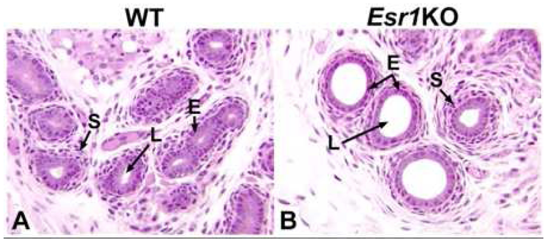
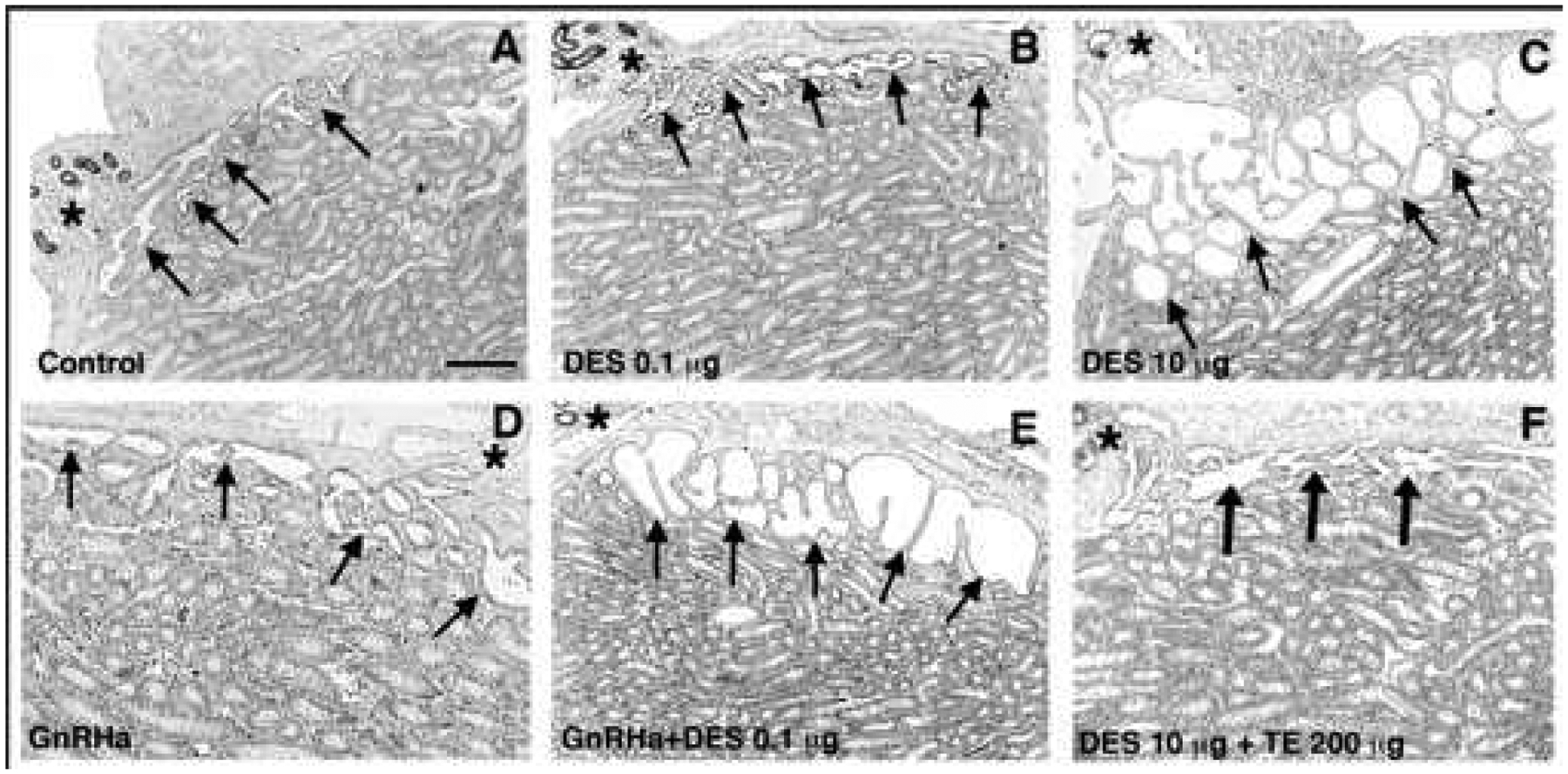
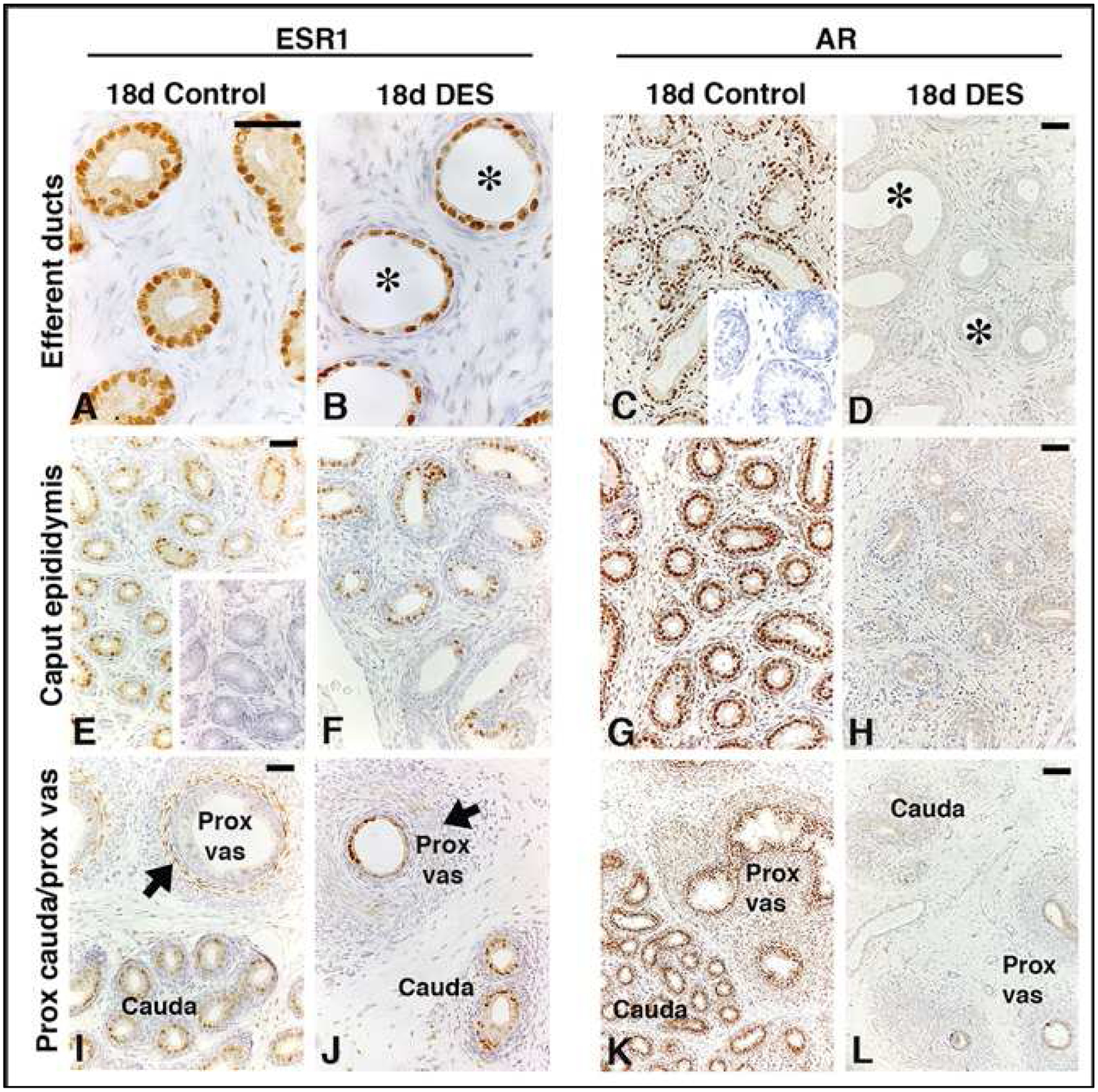
Estrogen has always been considered the female hormone and testosterone the male hormone. However, estrogen's presence in the testis and deleterious effects of estrogen treatment during development have been known for nearly 90 years, long before estrogen receptors (ESRs) were discovered. Eventually it was learned that testes actually synthesize high levels of estradiol (E2) and sequester high concentrations in the reproductive tract lumen, which seems contradictory to the overwhelming number of studies showing reproductive pathology following exogenous estrogen exposures. For too long, the developmental pathology of estrogen has dominated our thinking, even resulting in the "estrogen hypothesis" as related to the testicular dysgenesis syndrome. However, these early studies and the development of an Esr1 knockout mouse led to a deluge of research into estrogen's potential role in and disruption of development and function of the male reproductive system. What is new is that estrogen action in the male cannot be divorced from that of androgen. This paper presents what is known about components of the estrogen pathway, including its synthesis and target receptors, and the need to achieve a balance between androgen- and estrogen-action in male reproductive tract differentiation and adult functions. The review focuses on what is known regarding development of the male reproductive tract, from the rete testis to the vas deferens, and examines the expression of estrogen receptors and presence of aromatase in the male reproductive system, traces the evidence provided by estrogen-associated knockout and transgenic animal models and discusses the effects of fetal and postnatal exposures to estrogens. Hopefully, there will be enough here to stimulate discussions and new investigations of the androgen:estrogen balance that seems to be essential for development of the male reproductive tract.

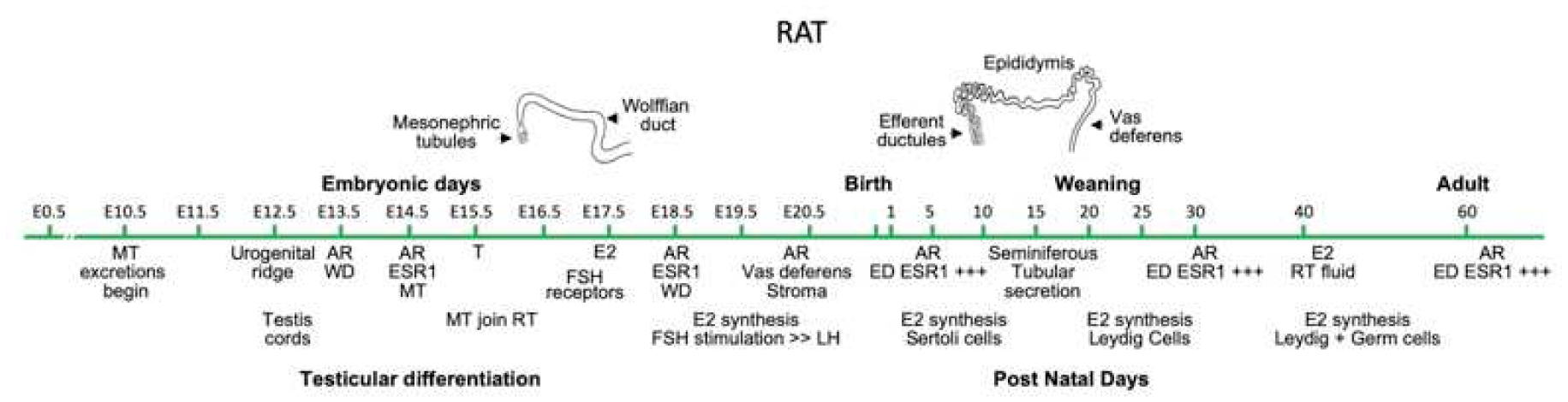
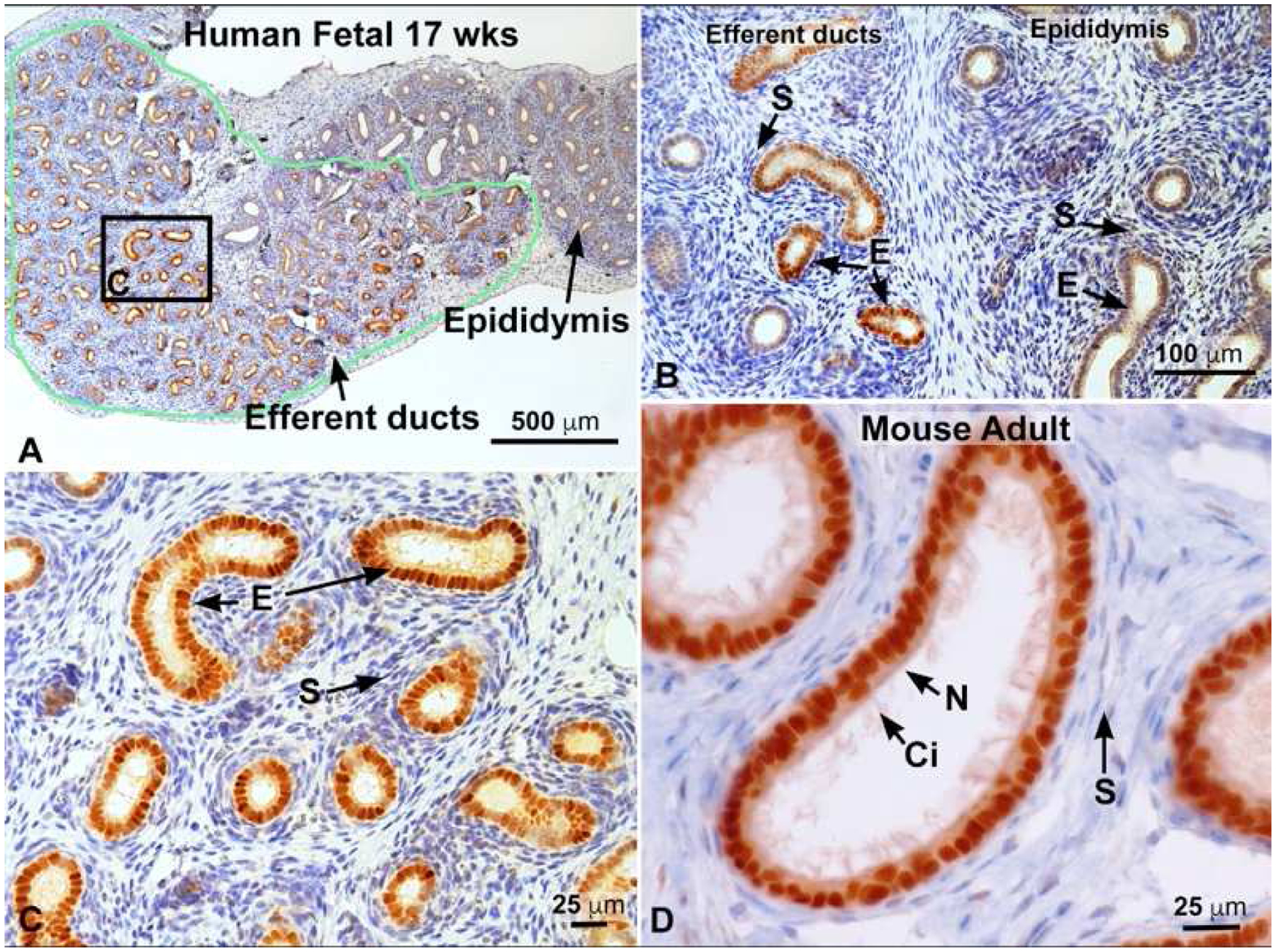
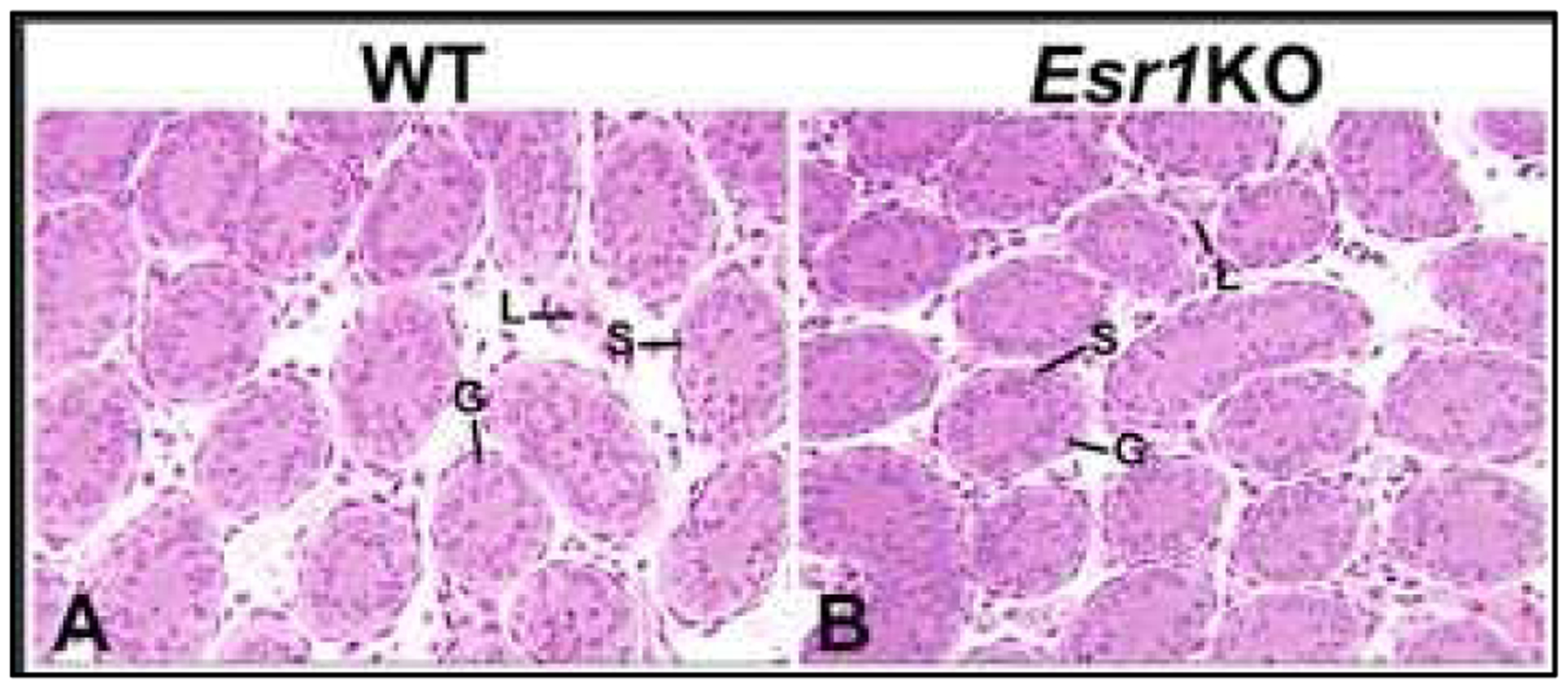
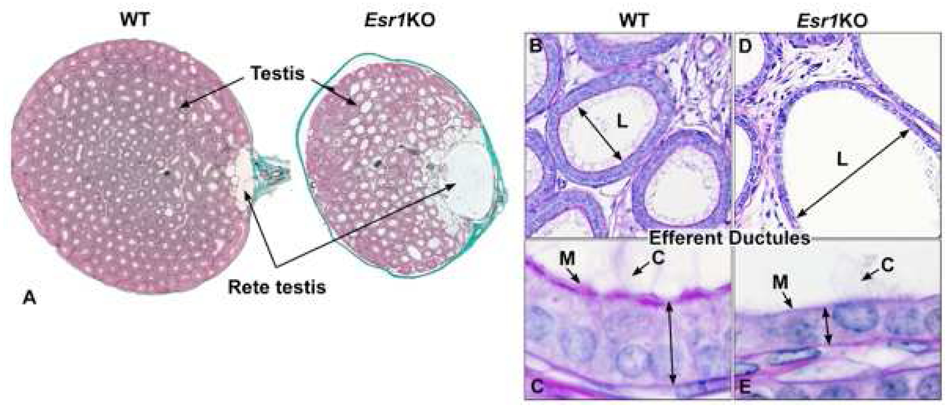
Keywords: Development; Differentiation; Efferent ductule; Environmental estrogens; Epididymis; Estrogen; Estrogen receptor; Fetal; Male reproduction; Mesonephric tubule; Mesonephros; Neonatal; Rete cord; Rete testis; Testicular dysgenesis syndrome; Testis; Vas deferens; Wolffian duct.


Conflict of interest statement

Competing interests statements:

Rex Hess has an appointment as Director of Science at Epivara, Inc., a start-up company dedicated to research and development of animal sterilization methods. Epivara provided financial assistance for the literature review but had no influence on the preparation of the manuscript.

Richard Sharpe has no competing interests to declare.

Barry Hinton has no competing interests to declare

Figures

Fig 1.
Fig 1.
Time course major events in development and differentiation of the rat male reproductive system. Although efferent ductules were emphysized for ESR1 expression, epididymal epithelium has positive expression in select cell types (see section 3). MT, mesonephric tubules; AR, androgen receptor; ESR, estrogen receptor; WD, Wolffian duct; T, testosterone; E2, estrogen; FSH, Follicle Stimulating Hormone; LH, Luteinizing Hormone; ED, efferent ductules; RT, rete testis. Adapted from Hinton & Avellar (2018).
Fig 2.
Fig 2.
Connections in the E15.5 mouse embryo showing testis, rete testis cord, mesonephric tubules and Wolffian duct. On the right is a three-dimensional model. Photos were modified from Combes et al. (2009) and De Mello Santos et al. (2019) and reproduced by permission of Wiley Press.
Fig. 3.
Fig. 3.
Estrogen receptor 1 (ESR1) immunostaining. A) 17 week fetal human efferent ductules in the head of the epididymis outlined in green and staining intensely for ESR1. To the right is the immature epididymal duct, which appears to have far less staining. B) Fetal 17 week human tissue showing efferent ductules to the left and epididymis to the right. Efferent duct stroma (S) is negative, but the epithelial cells (E) are strongly positive for ESR1, while epididymal epithelium is less positive and some stromal cells show slight staining. C) Higher magnification of area outlined in A. Efferent duct epithelial cells (E) are strongly positive for ESR1, but the stroma (S) is negative. D) Mouse adult efferent ductules showing intense staining for ESR1 in both nonciliated (N) resorptive epithelial cells, as well as the motile ciliated cells (Ci). Stromal (S) cells are mostly negative. Human fetal photos provided by Dr. Gerald Cunha, with approval by the Committee on Human Research at UCSF, IRB# 12–08813); Cunha et al. (2020).
Fig 4.
Fig 4.
Testes from PND6 wild-type (WT) and Esr1KO mice. A. WT seminiferous tubules are small in diameter with no lumen and an epithelium consisting of Sertoli and germ cells. Leydig cells are seen in the interstitial space. B. Seminiferous tubules and interstitial space in the Esr1KO mouse are similar to WT at this age and show no formation of a lumen.
Fig 5.
Fig 5.
Testis, rete testis and efferent ductules in the adult wild-type (WT) and Esr1KO mice. A) WT testis shows normal seminiferous tubular lumens with a small flattened rete testis where sperm exit to enter the efferent ductules. In contrast, the rete testis in an Esr1KO mouse shows excessive dilation. B) WT efferent ductules showing a normal luminal (L) diameter. C) Enlargement of the WT epithelium to illustrate normal height, long motile cilia (C) extending into the lumen and adjacent thick microvillus boarder (M) of the nonciliated, resorptive cells that are responsible for reabsorption of nearly 90% of the luminal fluids. D) Esr1KO efferent ductule, showing hyperdilation of the lumen (L). E) Higher magnification of the Esr1KO epithelium, which shows the dramatic decrease in height and loss of apical cytoplasm and microvilli (M) in the resorptive cells. Motile cilia (C) extend into the lumen, but appear to be fewer in number and shorter in length. Adapted from figures in Nanjappa et al. (2016) and Hess et al. (2011) with permission from Oxford University Press and John Wiley and Sons.
Fig 6.
Fig 6.
Efferent ductules from PND 6 wild-type (WT) and Esr1KO mice. A) WT ductules have small diameters with tiny, but open lumens (L). The epithelium is short but taller than in the Esr1KO and the surrounding stromal cells make up about two layers. B) Efferent ductules from the Esr1KO mouse show strong luminal (L) dilation at this early age and the epithelium (E) is already decreased in height. The stromal cells appear normal.
Fig. 7.
Fig. 7.
Effect of neonatal treatment with vehicle (control; A), a low dose (0.1 μg; B) or a high dose (10 μg; C) of diethylstilbestrol (DES), a GnRH antagonist (GnRHa; D), 0.1 μg DES plus GnRHa (E) or 10 μg DES + 200 μg testosterone esters (TE) (F) on the size of the rete testis lumen (arrows) at postnatal age 15 d. Note that all sections were obtained in the region in which the efferent ducts (asterisks) emerged from the rete along the testis capsule. Sections were immunostained for ESR1, hence the dark staining of nuclei of epithelial cells in the efferent ducts. Scale bar, 400 μm. Adapted from figures in Rivas et al (2002) and McKinnell et al (2001) with permission from Oxford University Press and John Wiley and Sons.
Fig. 8.
Fig. 8.
Effect of neonatal treatment of rats with vehicle (controls) or a high dose of diethylstilbestrol (DES; 10 μg) on immunoexpression of ESR1 (left-hand panels) and AR (right-hand panels) on postnatal day 18 in the efferent ductules (top row), caput epididymis (middle row) and proximal cauda/proximal vas deferens (bottom row). Note that, unlike in controls (A), epithelial expression of ESR1 in DES-treated rats extends into the proximal (J) regions of the vas deferens. Immunoexpression of ESR1 was unaffected in the efferent ducts (A vs. B), caput (E vs. F), and proximal cauda (I vs. J). Note also the reduction in height of the epithelium in the efferent ducts (A vs. B) and proximal vas deferens of DES-treated rats (I vs. J and K vs.L). DES also induced luminal distension of the efferent ducts (asterisks; A vs B). DES treatment profoundly reduced both epithelial and stromal expression of AR in the efferent ducts (C vs. D), caput epididymis (G vs. H) and proximal cauda and vas (K vs. L). Insets in panels C and E show negative controls in which the primary antibody was preabsorbed with the appropriate recombinant protein. Arrowheads point to stromal immunoexpression of ESR1 in the proximal vas (I and J). Scale bars, 50 μm. Adapted from figures in Atanassova et al 2001 and McKinnell et al (2001) with permission from Oxford University Press and John Wiley and Sons.

References

    1. Abot A, Fontaine C, Raymond-Letron I, Flouriot G, Adlanmerini M, Buscato M, Otto C, Berges H, Laurell H, Gourdy P, Lenfant F, Arnal JF, 2013. The AF-1 activation function of estrogen receptor alpha is necessary and sufficient for uterine epithelial cell proliferation in vivo. Endocrinology 154, 2222–2233. - PubMed
    1. Aceitero J, Llanero M, Parrado R, Pena E, Lopez-Beltran A, 1998. Neonatal exposure of male rats to estradiol benzoate causes rete testis dilation and backflow impairment of spermatogenesis. Anat Rec 252, 17–33. - PubMed
    1. Ahlbory-Dieker DL, Stride BD, Leder G, Schkoldow J, Trolenberg S, Seidel H, Otto C, Sommer A, Parker MG, Schutz G, Wintermantel TM, 2009. DNA binding by estrogen receptor-alpha is essential for the transcriptional response to estrogen in the liver and the uterus. Mol Endocrinol 23, 1544–1555. - PMC - PubMed
    1. Amann RP, Ganjam VK, 1976. Steroid production by the bovine testis and steroid transfer across the pampiniform plexus. Biol Reprod 15, 695–703. - PubMed
    1. Amano A, Kondo Y, Noda Y, Ohta M, Kawanishi N, Machida S, Mitsuhashi K, Senmaru T, Fukui M, Takaoka O, Mori T, Kitawaki J, Ono M, Saibara T, Obayashi H, Ishigami A, 2017. Abnormal lipid/lipoprotein metabolism and high plasma testosterone levels in male but not female aromatase-knockout mice. Archives of biochemistry and biophysics 622, 47–58. - PubMed

Publication types

MeSH terms