. 2021 Mar 10;95(7):e00014-21.
doi: 10.1128/JVI.00014-21. Epub 2021 Jan 13.

Non-steroidal anti-inflammatory drugs dampen the cytokine and antibody response to SARS-CoV-2 infection

Affiliations

Non-steroidal anti-inflammatory drugs dampen the cytokine and antibody response to SARS-CoV-2 infection

Jennifer S Chen et al. J Virol. .

Abstract

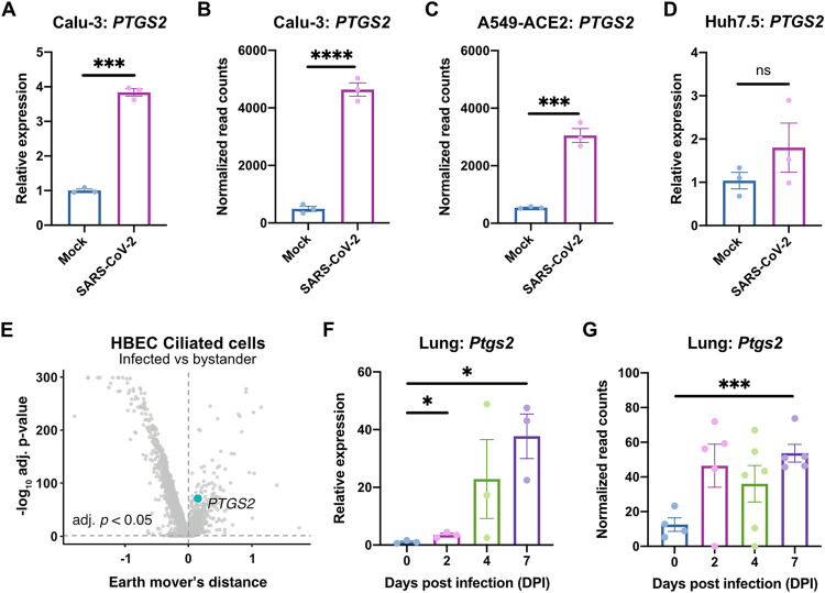
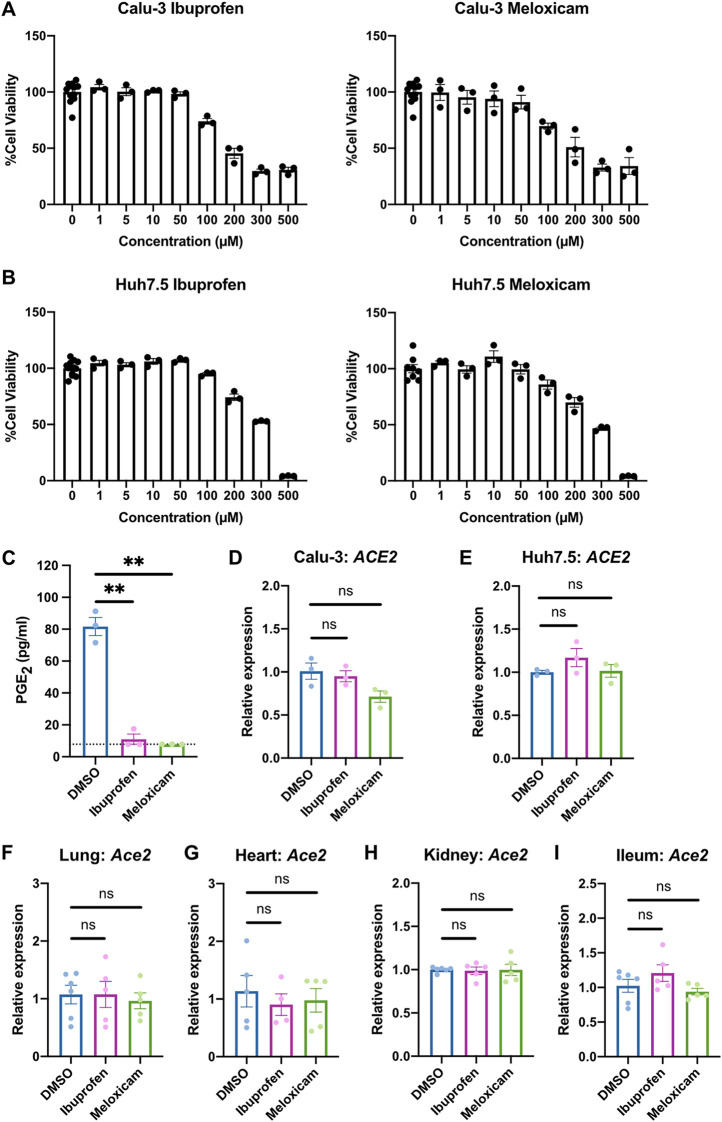
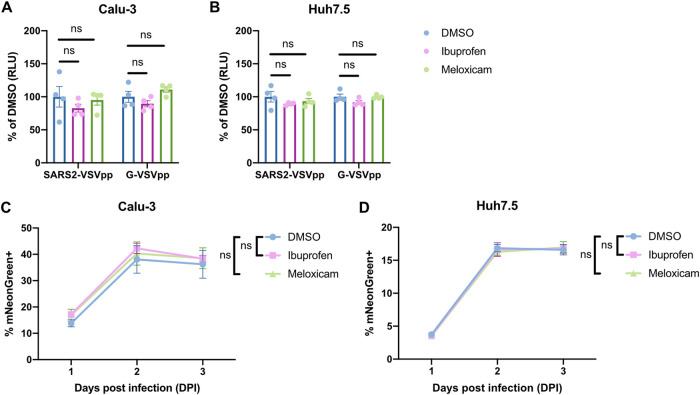
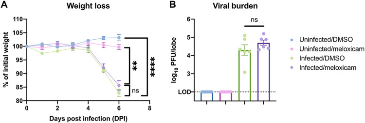
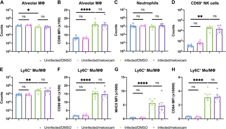
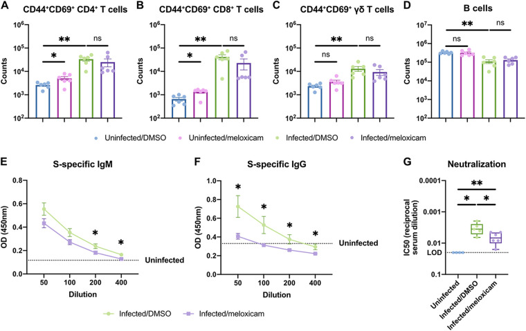
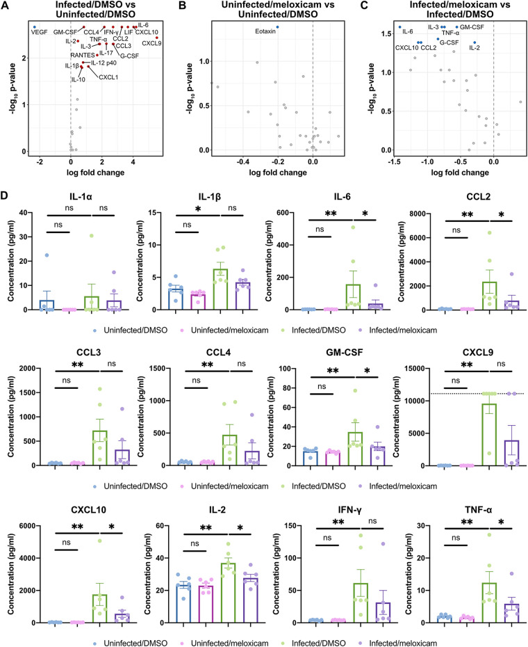
Identifying drugs that regulate severe acute respiratory syndrome coronavirus 2 (SARS-CoV-2) infection and its symptoms has been a pressing area of investigation during the coronavirus disease 2019 (COVID-19) pandemic. Nonsteroidal anti-inflammatory drugs (NSAIDs), which are frequently used for the relief of pain and inflammation, could modulate both SARS-CoV-2 infection and the host response to the virus. NSAIDs inhibit the enzymes cyclooxygenase-1 (COX-1) and cyclooxygenase-2 (COX-2), which mediate the production of prostaglandins (PGs). As PGs play diverse biological roles in homeostasis and inflammatory responses, inhibiting PG production with NSAIDs could affect COVID-19 pathogenesis in multiple ways, including: (1) altering susceptibility to infection by modifying expression of angiotensin-converting enzyme 2 (ACE2), the cell entry receptor for SARS-CoV-2; (2) regulating replication of SARS-CoV-2 in host cells; and (3) modulating the immune response to SARS-CoV-2. Here, we investigate these potential roles. We demonstrate that SARS-CoV-2 infection upregulates COX-2 in diverse human cell culture and mouse systems. However, suppression of COX-2 by two commonly used NSAIDs, ibuprofen and meloxicam, had no effect on ACE2 expression, viral entry, or viral replication. In contrast, in a mouse model of SARS-CoV-2 infection, NSAID treatment reduced production of pro-inflammatory cytokines and impaired the humoral immune response to SARS-CoV-2 as demonstrated by reduced neutralizing antibody titers. Our findings indicate that NSAID treatment may influence COVID-19 outcomes by dampening the inflammatory response and production of protective antibodies rather than modifying susceptibility to infection or viral replication.ImportancePublic health officials have raised concerns about the use of nonsteroidal anti-inflammatory drugs (NSAIDs) for treating symptoms of coronavirus disease 2019 (COVID-19). NSAIDs inhibit the enzymes cyclooxygenase-1 (COX-1) and cyclooxygenase-2 (COX-2), which are critical for the generation of prostaglandins - lipid molecules with diverse roles in homeostasis and inflammation. Inhibition of prostaglandin production by NSAIDs could therefore have multiple effects on COVID-19 pathogenesis. Here, we demonstrate that NSAID treatment reduced both the antibody and pro-inflammatory cytokine response to SARS-CoV-2 infection. The ability of NSAIDs to modulate the immune response to SARS-CoV-2 infection has important implications for COVID-19 pathogenesis in patients. Whether this occurs in humans and whether it is beneficial or detrimental to the host remains an important area of future investigation. This also raises the possibility that NSAIDs may alter the immune response to SARS-CoV-2 vaccination.


Figures

FIG 1
FIG 1
SARS-CoV-2 infection induces PTGS2 expression in human cells and mice. (A) Calu-3 cells were infected with SARS-CoV-2 at an MOI of 0.05. PTGS2 expression was measured at 2 dpi, normalized to ACTB. (B and C) PTGS2 expression in Calu-3 (B) and ACE2-overexpressing A549 (A549-ACE2) (C) cells following SARS-CoV-2 infection. The data are from GSE147507 (26). (D) Huh7.5 cells were infected with SARS-CoV-2 at an MOI of 0.05. PTGS2 expression was measured at 2 dpi, normalized to ACTB. (E) HBECs were cultured at an air-liquid interface and then infected at the apical surface with 104 PFU of SARS-CoV-2. Cells were collected at 1, 2, and 3 dpi for single-cell RNA sequencing (scRNA-seq) (28). A volcano plot of differentially expressed genes in infected versus bystander ciliated cells pooled from all time points is shown. PTGS2 is highlighted. (F) K18-hACE2 mice were infected intranasally with 1.2 × 106 PFU of SARS-CoV-2. Ptgs2 expression in the lung was measured at 0, 2, 4, and 7 dpi. (G) Ptgs2 expression in the lung of K18-hACE2 mice following intranasal SARS-CoV-2 infection. The data are from GSE154104 (36). All data points in this figure are presented as means ± the standard errors of the mean (SEM). Data were analyzed by Welch’s two-tailed, unpaired t test (A, D, and F); Student two-tailed, unpaired t test (B, C, and G); and two-sided Mann-Whitney U test with continuity and Benjamini-Hochberg correction (E). *, P < 0.05; ***, P < 0.001; ****, P < 0.0001. Data in panels A and D are representative of two independent experiments with three replicates per condition.
FIG 2
FIG 2
NSAID treatment does not affect ACE2 expression in human cells and mice. (A and B) Calu-3 (A) and Huh7.5 (B) cells were treated with different concentrations of ibuprofen or meloxicam for 48 h. Cell viability was measured and calculated as a percentage of no treatment. (C) Calu-3 cells were treated with DMSO, 50 μM ibuprofen, or 50 μM meloxicam for 48 h. The levels of prostaglandin E2 (PGE2) were measured in the supernatant. The dotted line represents the limit of detection. (D and E) Calu-3 (D) and Huh7.5 (E) cells were treated with DMSO, 50 μM ibuprofen, or 50 μM meloxicam for 24 h. ACE2 expression was measured and normalized to ACTB. (F to I) C57BL/6 mice were treated intraperitoneally with DMSO, 30 mg/kg ibuprofen, or 1 mg/kg meloxicam daily for 4 days. Ace2 expression was measured in the lung (F), heart (G), kidney (H), and ileum (I), normalized to Actb. All data points in this figure are presented as means ± the SEM. Data were analyzed by Welch’s two-tailed, unpaired t test (C to I). **, P < 0.01; ns, not significant. Data in panels A to E are representative of two independent experiments with three replicates per condition; data in panels F to I are pooled from two independent experiments with a total of four to six mice per condition.
FIG 3
FIG 3
NSAID treatment does not affect SARS-CoV-2 entry or replication in vitro. (A and B) Calu-3 (A) and Huh7.5 (B) cells were pretreated with DMSO, 50 μM ibuprofen, or 50 μM meloxicam for 24 h and then infected with SARS2-VSVpp or G-VSVpp expressing Renilla luciferase. Luminescence was measured at 24 h postinfection (hpi) and normalized to DMSO for each infection. (C and D) Calu-3 (C) and Huh7.5 (D) cells were pretreated with DMSO, 50 μM ibuprofen, or 50 μM meloxicam for 24 h and then infected with mNeonGreen reporter replication-competent SARS-CoV-2 (icSARS-CoV-2-mNG) at an MOI of 1. The frequency of infected cells was measured by mNeonGreen expression at 1, 2, and 3 dpi. All data points in this figure are presented as means ± the SEM. Data were analyzed by Student two-tailed, unpaired t test (A and B) and two-way ANOVA (C and D). ns, not significant. Data in panels A and B are representative of two independent experiments with four replicates per condition; data in panels C and D are representative of two independent experiments with five replicates per condition.
FIG 4
FIG 4
NSAID treatment does not affect SARS-CoV-2-induced weight loss or lung viral burden in mice. (A and B) K18-hACE2 mice were treated intraperitoneally with DMSO or 1 mg/kg meloxicam daily for 7 days starting 1 day prior to infection. K18-hACE2 mice were infected intranasally with 103 PFU of SARS-CoV-2 or left uninfected and monitored daily. (A) Weight change expressed as a percentage of initial weight. (B) Viral burden in the lungs at 6 dpi measured by plaque assay. All data points in this figure are presented as means ± the SEM. Data were analyzed by two-way ANOVA (A) and Student two-tailed, unpaired t test (B). **, P < 0.01; ****, P < 0.0001; ns, not significant. Data in panels A and B are pooled from two independent experiments with a total of six mice per condition.
FIG 5
FIG 5
NSAID treatment does not affect innate immune cell activation in the lungs of SARS-CoV-2-infected mice. (A to H) K18-hACE2 mice were treated intraperitoneally with DMSO or 1 mg/kg meloxicam daily for 7 days starting 1 day prior to infection. K18-hACE2 mice were infected intranasally with 103 PFU of SARS-CoV-2 or left uninfected. Flow cytometric analysis of the lungs at 6 dpi for alveolar macrophage (MΦ) counts (A) and expression of CD86 (B), neutrophil counts (C), activated CD69+ natural killer (NK) cell counts (D), and Ly6C+ monocyte/macrophage (Mo/MΦ) counts (E) and expression of CD86 (F), MHCII (G), and CD64 (H) results are shown. All data points in this figure are presented as means ± the SEM. Data were analyzed by two-tailed Mann-Whitney test (A to H). *, P < 0.05; **, P < 0.01; ****, P < 0.0001; ns, not significant. Data in panels A to H are pooled from two independent experiments with a total of six mice per condition.
FIG 6
FIG 6
NSAID treatment impairs systemic neutralizing antibody responses but not adaptive immune cell activation in the lungs of SARS-CoV-2-infected mice. (A to G) K18-hACE2 mice were treated intraperitoneally with DMSO or 1 mg/kg meloxicam daily for 7 days starting 1 day prior to infection. K18-hACE2 mice were infected intranasally with 103 PFU of SARS-CoV-2 or left uninfected. Flow cytometric analysis of the lungs at 6 dpi for activated CD44+CD69+ CD4+ T cells (A), CD44+ CD69+ CD8+ T cells (B), CD44+ CD69+ γδ T cells (C), and B cells (D) was performed. (E and F) Spike (S)-specific IgM (E) and IgG (F) titers in the serum at 6 dpi. (G) Neutralizing antibody titers in the serum at 6 dpi measured by SARS2-VSVpp pseudovirus neutralization assay. Data points in panels A to F are presented as means ± the SEM. Data points in panel G are presented as boxplots. Data were analyzed by two-tailed Mann-Whitney test (A to D, G) and Student two-tailed, unpaired t test (E and F). *, P < 0.05; **, P < 0.01; ns, not significant. Data in panels A to G are pooled from two independent experiments with a total of four to six mice per condition.
FIG 7
FIG 7
NSAID treatment dampens the induction of proinflammatory cytokines that are upregulated by SARS-CoV-2 infection in mice. (A to D) K18-hACE2 mice were treated intraperitoneally with DMSO or 1 mg/kg meloxicam daily for 7 days starting 1 day prior to infection. K18-hACE2 mice were infected intranasally with 103 PFU of SARS-CoV-2 or left uninfected. Cytokine levels were measured in lung homogenates at 6 dpi. (A to C) Volcano plots detailing the differential abundance of cytokines in lung homogenates from infected versus uninfected mice treated with DMSO (A), uninfected mice treated with meloxicam versus DMSO (B), and infected mice treated with meloxicam versus DMSO (C). Significantly upregulated (red) and downregulated (blue) cytokines are labeled. (D) Levels of proinflammatory cytokines in lung homogenates from uninfected mice treated with DMSO, uninfected mice treated with meloxicam, infected mice treated with DMSO, and infected mice treated with meloxicam. The dotted line represents the upper limit of quantification. Data points in panel D are presented as means ± the SEM. Data were analyzed by two-tailed Mann–Whitney test (A to D). *, P < 0.05; **, P < 0.01; ns, not significant. Data in panels A to D are pooled from two independent experiments with a total of six mice per condition. Additional cytokine data are shown in Fig. S2 in the supplemental material.

References

    1. Day M. 2020. Covid-19: ibuprofen should not be used for managing symptoms, say doctors and scientists. BMJ 368:m1086. - PubMed
    1. Powis S. 2020. Novel coronavirus: anti-inflammatory medications. Medicines and Healthcare Products Regulatory Agency, London, United Kingdom.
    1. Ricciotti E, FitzGerald GA. 2011. Prostaglandins and inflammation. Arterioscler Thromb Vasc Biol 31:986–1000. doi:10.1161/ATVBAHA.110.207449. - DOI - PMC - PubMed
    1. Zhou P, Yang X-L, Wang X-G, Hu B, Zhang L, Zhang W, Si H-R, Zhu Y, Li B, Huang C-L, Chen H-D, Chen J, Luo Y, Guo H, Jiang R-D, Liu M-Q, Chen Y, Shen X-R, Wang X, Zheng X-S, Zhao K, Chen Q-J, Deng F, Liu L-L, Yan B, Zhan F-X, Wang Y-Y, Xiao G-F, Shi Z-L. 2020. A pneumonia outbreak associated with a new coronavirus of probable bat origin. Nature 579:270–273. doi:10.1038/s41586-020-2012-7. - DOI - PMC - PubMed
    1. Fang L, Karakiulakis G, Roth M. 2020. Are patients with hypertension and diabetes mellitus at increased risk for COVID-19 infection? Lancet Respir Med 8:e21. doi:10.1016/S2213-2600(20)30116-8. - DOI - PMC - PubMed

LinkOut - more resources