. 2021 Jan 19;118(3):e2012935118.
doi: 10.1073/pnas.2012935118.

The 3- O-sulfation of heparan sulfate modulates protein binding and lyase degradation

Affiliations

The 3- O-sulfation of heparan sulfate modulates protein binding and lyase degradation

Pradeep Chopra et al. Proc Natl Acad Sci U S A. .

Abstract

Humans express seven heparan sulfate (HS) 3-O-sulfotransferases that differ in substrate specificity and tissue expression. Although genetic studies have indicated that 3-O-sulfated HS modulates many biological processes, ligand requirements for proteins engaging with HS modified by 3-O-sulfate (3-OS) have been difficult to determine. In particular, the context in which the 3-OS group needs to be presented for binding is largely unknown. We describe herein a modular synthetic approach that can provide structurally diverse HS oligosaccharides with and without 3-OS. The methodology was employed to prepare 27 hexasaccharides that were printed as a glycan microarray to examine ligand requirements of a wide range of HS-binding proteins. The binding selectivity of antithrombin-III (AT-III) compared well with anti-Factor Xa activity supporting robustness of the array technology. Many of the other examined HS-binding proteins required an IdoA2S-GlcNS3S6S sequon for binding but exhibited variable dependence for the 2-OS and 6-OS moieties, and a GlcA or IdoA2S residue neighboring the central GlcNS3S. The HS oligosaccharides were also examined as inhibitors of cell entry by herpes simplex virus type 1, which, surprisingly, showed a lack of dependence of 3-OS, indicating that, instead of glycoprotein D (gD), they competitively bind to gB and gC. The compounds were also used to examine substrate specificities of heparin lyases, which are enzymes used for depolymerization of HS/heparin for sequence determination and production of therapeutic heparins. It was found that cleavage by lyase II is influenced by 3-OS, while digestion by lyase I is only affected by 2-OS. Lyase III exhibited sensitivity to both 3-OS and 2-OS.

Keywords: 3-O-sulfation; anti-Factor Xa; glycan microarray; heparin lyases; herpes simplex virus 1.


Conflict of interest statement

The authors declare no competing interest.

Figures

Fig. 1.
Fig. 1.
(A) Identified substructures having 3-OS−bearing glucosamine. (B) General structure of target HS hexasaccharides, featuring structurally diverse sequence (shaded pyranose rings), site of sulfation (text in red), uronic acid composition (wavy bond at C-5 carboxylic acid), constant reducing end GlcN and nonreducing end disaccharide, and an anomeric linker for fabrication of HS arrays. (C) Hexasaccharides numbering and backbone composition, variable core trisaccharide in red color; NS, N-sulfate; 2S: 2-OS; 3S, 3-OS; 6S, 6-OS. (D) Disaccharide building blocks comprising acceptors 10 to 12 and donors 13 to 19 for modular assembly of hexasaccharides.
Fig. 2.
Fig. 2.
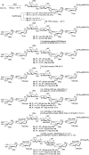
An example of the synthesis of hexasaccharides having central GlcNS modified by 3,6-OS (1A), 3-OS (1B), and 6-OS (1C). Synthetic schemes of remaining hexasaccharides are provided in SI Appendix, Schemes S3–S10. Reagents: TfOH, trifluoromethanesulfonic acid; CH2Cl2, dichloromethane; Et3N, triethylamine; EtOH, ethanol; DMF, dimethylformamide; LiOH, lithium hydroxide; H2O2, hydrogen peroxide; NaOH, sodium hydroxide; PMe3, trimethylphosphine; MeOH, methanol; Pd/C, palladium on carbon; H2, hydrogen gas; tBuOH/H2O, tert-butanol/water; Pd(OH)2/C, palladium hydroxide on carbon.
Fig. 3.
Fig. 3.
Structural analysis of hexasaccharides by NMR spectroscopy. (A) Structure of hexasaccharides with core trisaccharide GlcA-GlcNS-GlcA with variable 3- and/or 6-O-sulfation on central glucosamine. (B) Stacked plots showing 1H NMR of compounds 1A, 1B, and 1C; blue highlighted area, presence of 3-OS results in distinct downfield shift of GlcNH-3; green highlighted area, presence of 3-OS results in characteristic split and downfield shift of GlcNH-2. (C) The 1H-13C HSQC NMR of compound 1A; blue highlighted area, characteristic C-3 of 3-O-sulfated GlcN; green highlighted area, characteristic C-2 of 3-O-sulfated GlcN. (D) The 1H-1H–TOCSY NMR spectrum of compound 1A; characteristic correlation between ring protons (anomeric H-1, H-2, and H-3 protons) of 3-OS−bearing central GlcN.
Fig. 4.
Fig. 4.
Screening various HS recognizing proteins for their binding against an array of synthetic hexasaccharides printed at 100 μM concentration in a replicate of six. (A) AT-III/Human serpinC1 (10 μg/mL). (B) HC-II/Human serpinD1 (3 μg/mL). (C) Human FGF-7 (0.3 μg/mL). (D) Human FGF-9 (0.1 μg/mL). (E) Human BMP-2 (1 μg/mL). (F) Human FGFR-1/CD331 (10 μg/mL). (G) RAGEWRAGE RAGE/advanced glycosylation end product AGER (1 μg/mL). (H) Human stabilin-2/Stab-2 (30 μg/mL). (I) Mouse Nrp-1 (30 μg/mL). List of protein source (type of fusion tag, if present), primary antibodies, and secondary antibodies (type of fluorophore, if present) screened against HS microarray are presented in SI Appendix, Table S1. For dose–response of various HS recognizing protein screened for their binding to HS microarray, see SI Appendix Fig. S2. Data are presented as mean ± SD (n = 4). Representative data are shown for each protein, which was repeated at least three times.
Fig. 5.
Fig. 5.
Anticoagulant activity of hexasaccharides. (A) Factor Xa inhibition assays, HS hexamers were screened at concentrations 10 and 3 nM, respectively, with Fondaparinux (Fpx.) as positive control; higher concentration screening data are presented in SI Appendix, Fig. S3, symbol structures of identified hits are presented, and the variable core region is highlighted in blue. (B) Determination of IC50 of 4A, 4B, 7A, and 7B; an overlay of inhibition curve is presented, and individual inhibition curves are presented in SI Appendix, Fig. S4. (C) Table for IC50 value of identified hits. Symbol nomenclature for HS backbone monosaccharides is presented in the dashed box. Data are presented as mean ± SD (for A and B, n = 3). All experiments were performed three times at the minimum. R = O(CH2)5NH2.
Fig. 6.
Fig. 6.
Inhibition of HSV-1 infection of HCEs (RCB1834 HCE-T) by HS hexasaccharides. (A) Screening of hexasaccharides at 100 μg/mL, untreated and infected blank as positive control, and Fpx. was used as a 3-OS standard; box highlighted area shows HS oligosaccharides that elicited more than 80% reduction in virus infection; symbol structure of the hexamers and influence of sulfation site on viral infection; data presented are an average of three independent experiments. (B) Determination of IC50 values of 3,6-OS hits (7A, 8A, and 9A); 6-OS hits (7C, 8C, and 9C) are in SI Appendix, Fig. S5A. (C) Table of IC50 value of identified hits, 7A, 7C, 8A, 8C, 9A, and 9C. (D) GFP reporter HSV-1 infection assays; images for compound 7B, 8B, 8C, and 9B are in SI Appendix, Fig. S5B. (Scale bar, 100 μm.) Symbol nomenclature for HS backbone monosaccharides is presented in dashed box. Data are presented as mean ± SD (for A and B, n = 3). All experiments were performed at least as three independent experiments. R = O(CH2)5NH2.
Fig. 7.
Fig. 7.
Heparin lyases substrate requirements for the digestion of 3-OS−bearing HS. Mode of lyase cleavage: Lyase can cleave intact hexasaccharide (HFull) at site A—nonreducing end (NRE) uronic acid—and/or site B—reducing end (RE) uronic acid—to release different tetrasaccharides (TRE and TNRE) and disaccharides (DNRE, DINR, and DRE, where INR stands for inner disaccharide). Symbol nomenclature for HS backbone monosaccharides is presented in the dashed box. For illustration purposes, HS modifications other than 3-OS were not shown in the model substrate (HFull).
Fig. 8.
Fig. 8.
Summary figure of heparin lyase (I, II, and III) activities. Cleavage site color code: Blue rectangle represents cleavage at site A, orange rectangle represents cleavage at site B, green rectangle represents cleavage at both sites A and B, and red rectangle represents resistant substrates with no cleavage. (A) Hexasaccharides having GlcA toward the reducing end of central GlcN. (B) Hexasaccharides with IdoA toward reducing end of central GlcN. (C) Hexasaccharides with IdoA2S toward reducing end of central GlcN. Variable trisaccharide core UA ± 2S-GlcNS ± 6S ± 3S-UA ± 2S is highlighted in light blue.
Fig. 9.
Fig. 9.
Heparin lyases (I, II, and III) mode of action: Influence of 3-O-sulfation and uronic acid at potential cleavage site on HS hexamers digestion.

Similar articles

Cited by

References

    1. Sarrazin S., Lamanna W. C., Esko J. D., Heparan sulfate proteoglycans. Cold Spring Harb. Perspect. Biol. 3, a004952 (2011). - PMC - PubMed
    1. Esko J. D., Selleck S. B., Order out of chaos: Assembly of ligand binding sites in heparan sulfate. Annu. Rev. Biochem. 71, 435–471 (2002). - PubMed
    1. Bishop J. R., Schuksz M., Esko J. D., Heparan sulphate proteoglycans fine-tune mammalian physiology. Nature 446, 1030–1037 (2007). - PubMed
    1. Xu D., Esko J. D., Demystifying heparan sulfate-protein interactions. Annu. Rev. Biochem. 83, 129–157 (2014). - PMC - PubMed
    1. Sasisekharan R., Venkataraman G., Heparin and heparan sulfate: Biosynthesis, structure and function. Curr. Opin. Chem. Biol. 4, 626–631 (2000). - PubMed

Publication types

MeSH terms