. 2021 Jan 13;11(1):854.
doi: 10.1038/s41598-020-72125-0.

Color-tuning of natural variants of heliorhodopsin

Affiliations

Color-tuning of natural variants of heliorhodopsin

Se-Hwan Kim et al. Sci Rep. .

Abstract

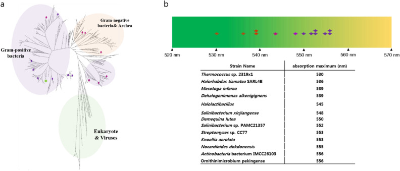
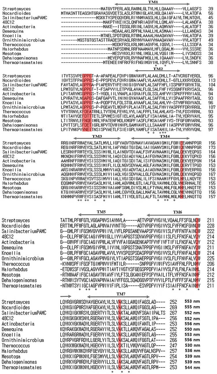
Microbial rhodopsins are distributed through many microorganisms. Heliorhodopsins are newly discovered but have an unclear function. They have seven transmembrane helices similar to type-I and type-II rhodopsins, but they are different in that the N-terminal region of heliorhodopsin is cytoplasmic. We chose 13 representative heliorhodopsins from various microorganisms, expressed and purified with an N-terminal His tag, and measured the absorption spectra. The 13 natural variants had an absorption maximum (λmax) in the range 530-556 nm similar to proteorhodopsin (λmax = 490-525 nm). We selected several candidate residues that influence rhodopsin color-tuning based on sequence alignment and constructed mutants via site-directed mutagenesis to confirm the spectral changes. We found two important residues located near retinal chromophore that influence λmax. We also predict the 3D structure via homology-modeling of Thermoplasmatales heliorhodopsin. The results indicate that the color-tuning mechanism of type-I rhodopsin can be applied to understand the color-tuning of heliorhodopsin.


Conflict of interest statement

The authors declare no competing interests.

Figures

Figure 1
Figure 1
Phylogenetic tree and distribution of absorption maxima for various heliorhodopsins. (a) The phylogenetic tree is built with ~ 474 heliorhodopsins. Thirteen of them were selected from various microorganisms: Thermococcus sp. 2,319 × 1 (530 nm), Halorhabdus tiamatea SARL4B (536 nm), Mesotoga infera (539 nm), Degalogenimonas alkenigignes (539 nm), Halolactibacillus (545 nm), Salinibacterium xinjiangense (548 nm), Demiquina lutea (550 nm), Salinibacterium sp. PAMC21357 (552 nm), Streptomyces sp. CC77 (553 nm), Knoellia aerolata (553 nm), Nocardioides dokdonensis (555 nm), Actinobacteria bacterium IMCC26103 (556 nm), Orinithinimicrobium pekingense (556 nm). The arrows indicate the position of heliorhodopsins listed above, and the green circle is 48C12 HeR. Heliorhodopsins with λmax > 550 nm are marked in purple. The other heliorhodopsins are marked with pink arrows. There is a tendency for heliorhodopsins λmax > 550 nm exhibit close genetic distance. (b) Distribution of λmax values of various heliorhodopsins. Thirteen heliorhodopsin proteins were purified. 48C12 HeR is marked on the light spectrum. The color of the diamond represents the λmax of each HeR. In nature, the variation in λmax for heliorhodopsin is almost ~ 30 nm.
Figure 2
Figure 2
Sequence alignment of 13 heliorhodopsin sequences. Four heliorhodopsins have an absorption maximum < 540 nm, one heliorhodopsin has an absorption maximum at 544 nm, and the other heliorhodopsins have absorption maxima > 550 nm. Significantly different amino acid regions are marked in red. The asterisk (*) indicates residues that comprise the retinal binding pocket.
Figure 3
Figure 3
Candidates for the residues involved in the color-tuning of natural variants of heliorhodopsins. The topology prediction of Ornithinimicrobium heliorhodopsin (λmax = 556 nm) obtained with Protter. Red open circles are candidate amino acids for the spectral tuning site. The comparison is the sequence from Fig. 2 (G59 ~ G62, Q159M, Q224M, and A254S of Ornithinimicrobium heliorhodopsin) and Thermococcus sp. 2,319 × 1 heliorhodopsin (F44 ~ A47, M145, M205, and S232 are corresponding residues). Mutants are designed to exchange candidate residues between Thermococcus heliorhodopsin and Ornithinimicrobium heliorhodopsin.
Figure 4
Figure 4
Absorption spectrum of N-terminal His-tagged Thermococcus heliorhodopsin (NTH) and N-terminal His-tagged Ornithinimicrobium heliorhodopsin (NOH). (a) The maximum absorption of WT and mutant NTH is between 530 and 542 nm. The double mutant (M205Q/S232A) showed a large shift to red (12 nm). The WT NTH absorption spectrum is shown as a dotted-line in the mutant absorption spectrum. (b) The maximum absorption of WT and mutant NOH is between 543 and 556 nm. The double mutant (Q224M/A251S) showed a large shift to blue (12 nm). The WT NOH absorption spectrum is shown as a dotted-line in the mutant absorption spectrum. (c) Table of λmax values for each WT and mutant heliorhodopsin.
Figure 5
Figure 5
Local view of the residues involved in spectral tuning in N-terminal His-tagged Thermococcus heliorhodopsin (NTH) and N-terminal His-tagged Ornithinimicrobium heliorhodopsin (NOH) with computer modeling. The distance on both sides of the retinal chromophore (Schiff base side and β-ionone ring side) is changed along with mutant. For the NOH, the distance between β-ionone and Gln224 is greater when the Gln is replaced with Met. However, the distance of the Schiff base side of the chromophore becomes shorter when Ala is replaced with Ser. In the case of NTH, the prediction is reversed. Chromophore retinal is green, the amino group is blue, the O atom of COO- is red, the C atoms of amino acids are cyan, and S-methyl is yellow.

References

    1. Spudich JL, Yang C-S, Jung K-H, Spudich EN. Retinylidene proteins: Structures and functions from archaea to humans. Annu. Rev. Cell Dev. Biol. 2000;16:365–392. doi: 10.1146/annurev.cellbio.16.1.365. - DOI - PubMed
    1. Engqvist MKM, et al. Directed evolution of gloeobacter violaceus rhodopsin spectral properties. J. Mol. Biol. 2015;427:205–220. doi: 10.1016/j.jmb.2014.06.015. - DOI - PubMed
    1. Zhang F, et al. The microbial opsin family of optogenetic tools. Cell. 2011;147:1446–1457. doi: 10.1016/j.cell.2011.12.004. - DOI - PMC - PubMed
    1. Zhang F, et al. Multimodal fast optical interrogation of neural circuitry. Nature. 2007;446:633–639. doi: 10.1038/nature05744. - DOI - PubMed
    1. McIsaac RS, Bedbrook CN, Arnold FH. Recent advances in engineering microbial rhodopsins for optogenetics. Curr. Opin. Struct. Biol. 2015;33:8–15. doi: 10.1016/j.sbi.2015.05.001. - DOI - PMC - PubMed

Publication types

LinkOut - more resources