Cerebral magnetic resonance imaging of coincidental infarction and small vessel disease in retinal artery occlusion
- PMID: 33441709
- PMCID: PMC7806736
- DOI: 10.1038/s41598-020-80014-9
Cerebral magnetic resonance imaging of coincidental infarction and small vessel disease in retinal artery occlusion
Abstract
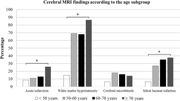
There are several reports in the literature on the association between non-arteritic retinal artery occlusion (NA-RAO) and acute ischemic stroke. We investigated the burden of small vessel disease (SVD) and cerebral coincident infarction observed on cerebral magnetic resonance imaging (MRI) in patients with newly diagnosed NA-RAO. In this retrospective, observational, case-series study, consecutive patients with NA-RAO who underwent cerebral MRI within one month of diagnosis between September 2003 and October 2018 were included. The classification of NA-RAO was based on ophthalmologic and systemic examinations. We also investigated the co-incident infarction and burden of underlying SVD, which were categorized as white matter hyperintensity lesion (WMH), cerebral microbleeds (CMB), and silent lacunar infarction (SLI). Among the 272 patients enrolled in the study, 18% presented co-incident infarction and 73% had SVD, which included WMH (70%), CMB (14%), and SLI (30%). Co-incident infarction, WMH, and SLI significantly increased with age: co-incident infarction was observed in 8% of young (< 50 years) patients and 30% of old (≥ 70 years) patients. The embolic etiology of RAO (large artery atherosclerosis, cardioembolism, and undetermined etiology) was significantly associated with the prevalence of SVD (82%: 70%: 64%, P = 0.002) and co-incident infarction (30%: 19%: 8%; P = 0.009). Therefore, high co-incidence of acute cerebral infarction and underlying SVD burden warrant careful neurologic examination and appropriate brain imaging, followed by management of NA-RAO. Urgent brain imaging is particularly pertinent in elderly patients with NA-RAO.
Conflict of interest statement
The authors declare no competing interests.
Figures


Similar articles
-
Collateral Recruitment Is Impaired by Cerebral Small Vessel Disease.Stroke. 2020 May;51(5):1404-1410. doi: 10.1161/STROKEAHA.119.027661. Epub 2020 Apr 6. Stroke. 2020. PMID: 32248770
-
Interhemispheric characterization of small vessel disease imaging markers after subcortical infarct.Brain Behav. 2016 Nov 3;7(1):e00595. doi: 10.1002/brb3.595. eCollection 2017 Jan. Brain Behav. 2016. PMID: 28127514 Free PMC article.
-
The presence and severity of cerebral small vessel disease increases the frequency of stroke in a cohort of patients with large artery occlusive disease.PLoS One. 2017 Oct 9;12(10):e0184944. doi: 10.1371/journal.pone.0184944. eCollection 2017. PLoS One. 2017. PMID: 28991905 Free PMC article.
-
Thrombolysis in acute stroke patients with cerebral small vessel disease.Cerebrovasc Dis. 2014;37(1):5-13. doi: 10.1159/000356796. Epub 2013 Dec 17. Cerebrovasc Dis. 2014. PMID: 24355873 Review.
-
[White Matter Lesion and Alzheimer's Disease: The Association between Small Vessel Disease and Neuropsychiatric Symptoms in Alzheimer's Disease].Brain Nerve. 2015 Apr;67(4):427-32. doi: 10.11477/mf.1416200158. Brain Nerve. 2015. PMID: 25846591 Review. Japanese.
Cited by
-
Central and Branch Retinal Artery Occlusion-Do They Harbor the Same Risk of Further Ischemic Events?J Clin Med. 2021 Jul 13;10(14):3093. doi: 10.3390/jcm10143093. J Clin Med. 2021. PMID: 34300257 Free PMC article.
-
Retinal Artery Occlusion and Its Impact on the Incidence of Stroke, Myocardial Infarction, and All-Cause Mortality during 12-Year Follow-Up.J Clin Med. 2022 Jul 14;11(14):4076. doi: 10.3390/jcm11144076. J Clin Med. 2022. PMID: 35887842 Free PMC article.
-
Prevalence and risk factors of ischemic monocular vision loss and concurrent brain ischemia.Eur Stroke J. 2023 Dec;8(4):982-988. doi: 10.1177/23969873231191577. Epub 2023 Aug 18. Eur Stroke J. 2023. PMID: 37593943 Free PMC article.
-
Central retinal artery occlusion or retinal stroke: a neurosonologist's perspective.Front Neurol. 2024 Jun 10;15:1397751. doi: 10.3389/fneur.2024.1397751. eCollection 2024. Front Neurol. 2024. PMID: 38915799 Free PMC article.
-
Redefining the Koizumi model of mouse cerebral ischemia: A comparative longitudinal study of cerebral and retinal ischemia in the Koizumi and Longa middle cerebral artery occlusion models.J Cereb Blood Flow Metab. 2022 Nov;42(11):2080-2094. doi: 10.1177/0271678X221109873. Epub 2022 Jun 24. J Cereb Blood Flow Metab. 2022. PMID: 35748043 Free PMC article.
References
-
- Chawla JC. Traumatic central retinal artery occlusion. Trans. Ophthalmol. Soc. U. K. 1972;92:777–784. - PubMed
Publication types
MeSH terms
LinkOut - more resources
Full Text Sources
Other Literature Sources