. 2021 Jan 13;11(1):967.
doi: 10.1038/s41598-020-79762-5.

Colony spreading of the gliding bacterium Flavobacterium johnsoniae in the absence of the motility adhesin SprB

Affiliations

Colony spreading of the gliding bacterium Flavobacterium johnsoniae in the absence of the motility adhesin SprB

Keiko Sato et al. Sci Rep. .

Abstract

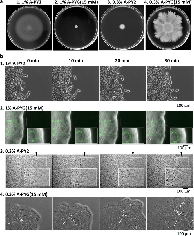
Colony spreading of Flavobacterium johnsoniae is shown to include gliding motility using the cell surface adhesin SprB, and is drastically affected by agar and glucose concentrations. Wild-type (WT) and ΔsprB mutant cells formed nonspreading colonies on soft agar, but spreading dendritic colonies on soft agar containing glucose. In the presence of glucose, an initial cell growth-dependent phase was followed by a secondary SprB-independent, gliding motility-dependent phase. The branching pattern of a ΔsprB colony was less complex than the pattern formed by the WT. Mesoscopic and microstructural information was obtained by atmospheric scanning electron microscopy (ASEM) and transmission EM, respectively. In the growth-dependent phase of WT colonies, dendritic tips spread rapidly by the movement of individual cells. In the following SprB-independent phase, leading tips were extended outwards by the movement of dynamic windmill-like rolling centers, and the lipoproteins were expressed more abundantly. Dark spots in WT cells during the growth-dependent spreading phase were not observed in the SprB-independent phase. Various mutations showed that the lipoproteins and the motility machinery were necessary for SprB-independent spreading. Overall, SprB-independent colony spreading is influenced by the lipoproteins, some of which are involved in the gliding machinery, and medium conditions, which together determine the nutrient-seeking behavior.


Conflict of interest statement

The authors declare no competing interests.

Figures

Figure 1
Figure 1
Colony spreading of WT F. johnsoniae on media containing different concentrations of agar and glucose. (a) Images of a colony spreading in a 9-cm diameter dish (5 days). Panel 1, 1% A-PY2; panel 2, 1% A-PY2 containing 15 mM glucose (1% A-PYG (15 mM)); panel 3, 0.3% A-PY2; panel 4, 0.3% A-PYG (15 mM). Colonies spread well on 1% A-PY2 and 0.3% A-PYG (15 mM). (b) Effect of agar and glucose concentrations on the behavior of bacteria in a spreading WT colony. Cells expressing GFP in their cytoplasm were added to the inoculated bacterial solution at a concentration of 1%, and movement of the bacterial cells at the colony edge was monitored. Images were recorded by fluorescence microscopy at 30-s intervals for 30 min. Panel 1, 1% A-PY2; panel 2, 1% A-PYG (15 mM); panel 3, 0.3% A-PY2; panel 4, 0.3% A-PYG (15 mM). At the leading edge of a spreading colony on 1% A-PY2 (panel 1), a small cell cluster moved outward toward the tip of a branch (Fig. S2a). By contrast, in the tip regions at the spreading front on 0.3% A-PYG (15 mM) (panel 4), windmill-like, flat structures including periodic stripes were carried outwards (Fig. S2d1). Time is shown at the top. The subpanels in row 2 and 3 are enlarged images of the regions marked by white rectangles. Arrowheads indicate the position of the leading edge of the colony in row 3. See Fig. S2 for selected frames showing the edge of each bacterial colony during 10 min of video capture.
Figure 2
Figure 2
Colony spreading of WT and ΔsprB mutant F. johnsoniae on 0.3% A-PYG (15 mM). (a) Optical microscopy image. The branching pattern of the ΔsprB colony was less complex than that of the WT. (b) Spreading colonies of F. johnsoniae immersed in aqueous glucose buffer imaged by ASEM. The colonies were aldehyde-fixed and stained using the NCMIR method. The top of the biofilm was observed. Both WT and ΔsprB mutant cells were buried in the extracellular matrix covering the top of the colony, reflecting biofilm formation. Upper panels, WT; lower panels, ΔsprB mutant.
Figure 3
Figure 3
TEM of WT and ΔsprB mutant F. johnsoniae spreading colonies on 0.3% A-PYG (15 mM). (a–d) Colonies were embedded in Epon resin, sliced into 70-nm sections, and observed by TEM. Sections of WT (a) and ΔsprB (b) colonies are shown. The cells were interspersed within the intercellular matrix. (c,d) High magnification images of a WT colony (c) and a ΔsprB colony (d). In both cases, the left and right panels are images of the upper and lower regions of the spreading colony, respectively.
Figure 4
Figure 4
Higher magnification images of the spreading colonies shown in Fig. 3. (a) TEM image of a 400 nm-thick, unstained, Epon section of a WT colony spreading on 0.3% A-PYG (15 mM). The space between bacterial cells is occupied by fibers and vesicles (upper panels). Migration of bacterial cells into the 0.3% agar layer (lower panels). WT cells metastasize into the agar medium (arrowhead). (b,c) Grid-stamp images of the spreading front region of WT (b) and ΔsprB (c) colonies.
Figure 5
Figure 5
SDS-PAGE analysis of the lipoproteins essential for colony spreading. (a) Comparison of the cell surface proteins of WT, ΔsprB and gldK F. johnsoniae on 0.3, 0.5 and 1% A-PYG (15 mM). Protein bands 1–16 were analyzed by peptide mass fingerprinting. A greater abundance of lipoproteins was present under colony expansion conditions on 0.3% A-PYG (15 mM) than under non-expansion conditions on 1% A-PYG (15 mM). (b) Comparison of the cell surface proteins of WT, ΔlolA, Δcsl, ΔsprB, ΔsprB ΔlolA and ΔsprB Δcsl F. johnsoniae on 0.3% A-PYG (15 mM). Protein bands 17–28 were analyzed by peptide mass fingerprinting. (c) Identification of protein bands present in the SDS-PAGE gel shown in sections a (band nos. 1–16) and b (band nos. 17–28) using mass spectrometry. (d) Colony spreading of WT, Δcsl, ΔsprB and ΔsprB Δcsl F. johnsoniae on 1% A-PY2 and 0.3% A-PYG (15 mM).
Figure 6
Figure 6
TEM of Δcsl mutant F. johnsoniae spreading colonies on 0.3% A-PYG (15 mM). Colonies were embedded in Epon resin, sliced into 70-nm serial sections, and observed by TEM. (a) Sections of Δcsl F. johnsoniae colonies. Similar to the WT and ΔsprB, the cells were interspersed within the intercellular matrix. (b) TEM image of a 400 nm-thick, unstained, Epon section of a Δcsl mutant colony spread on 0.3% A-PYG (15 mM). The intercellular space between bacterial cells was occupied by fibers and vesicles. (c) Higher magnification images of the squares in (b). Again, similar to WT and ΔsprB, the intercellular matrix was occupied by many intertwined fibers and secreted vesicles, suggesting biofilm formation.
Figure 7
Figure 7
ASEM images of F. johnsoniae. Cells were cultured in CYE (casitone-yeast extract) liquid medium directly on the SiN film of an ASEM dish, fixed with paraformaldehyde and glutaraldehyde, and stained as described in the methods section. The cells were immersed in glucose solution and observed using ASEM. (a) WT, (b) Δcsl, (c) ΔlolA. Upper panels, positively charged Nanogold-labeled cells; lower panels, Nanogold-labeled cells counterstained by the NCMIR method to visualize vesicles. Both the outer membranes and filaments 200 nm in length were clearly imaged for all of the cells. Scale bar 1 μm. (d) The polulation density of vesicles (vesicles/100 μm2) was measured. More vesicles were released from Δcsl cells than from WT cells (Fig. S8). (e) The area of vesicles (nm2) was measured. Vesicles released from Δcsl cells were almost the same size as vesicles released by the WT. The error bars on the graph correspond to the SD.
Figure 8
Figure 8
Grid stamp images of the edge of Δcsl mutant F. johnsoniae spreading colonies on 0.3% A-PYG (15 mM). Left: grid-stamp images of bacteria in the spreading front region. Right: high magnification image of the squares indicated in the left panels. Many vesicles were released from the Δcsl cells. The dark spots of high electron density observed in the WT cells on 1% A-PY2 were not observed in Δcsl cells on 0.3% A-PYG (15 mM).

Similar articles

Cited by

References

    1. Hunnicutt DW, Kempf MJ, McBride MJ. Mutations in Flavobacterium johnsoniae gldF and gldG disrupt gliding motility and interfere with membrane localization of GldA. J. Bacteriol. 2002;184:2370–2378. doi: 10.1128/JB.184.9.2370-2378.2002. - DOI - PMC - PubMed
    1. Hunnicutt DW, McBride MJ. Cloning and characterization of the Flavobacterium johnsoniae gliding-motility genes gldB and gldC. J. Bacteriol. 2000;182:911–918. doi: 10.1128/JB.182.4.911-918.2000. - DOI - PMC - PubMed
    1. McBride MJ, Braun TF. GldI is a lipoprotein that is required for Flavobacterium johnsoniae gliding motility and chitin utilization. J. Bacteriol. 2004;186:2295–2302. doi: 10.1128/JB.186.8.2295-2302.2004. - DOI - PMC - PubMed
    1. Braun TF, McBride MJ. Flavobacterium johnsoniae GldJ is a lipoprotein that is required for gliding motility. J. Bacteriol. 2005;187:2628–2637. doi: 10.1128/JB.187.8.2628-2637.2005. - DOI - PMC - PubMed
    1. Braun TF, Khubbar MK, Saffarini DA, McBride MJ. Flavobacterium johnsoniae gliding motility genes identified by mariner mutagenesis. J. Bacteriol. 2005;187:6943–6952. doi: 10.1128/JB.187.20.6943-6952.2005. - DOI - PMC - PubMed

Publication types

MeSH terms

Supplementary concepts

LinkOut - more resources