. 2021 Jan 13;11(1):1027.
doi: 10.1038/s41598-020-79734-9.

Berberine-releasing electrospun scaffold induces osteogenic differentiation of DPSCs and accelerates bone repair

Affiliations

Berberine-releasing electrospun scaffold induces osteogenic differentiation of DPSCs and accelerates bone repair

Lan Ma et al. Sci Rep. .

Abstract

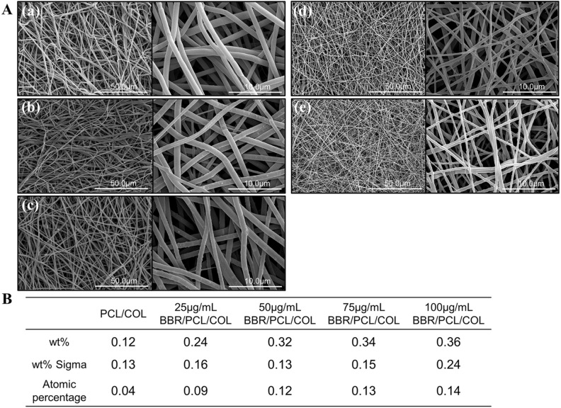
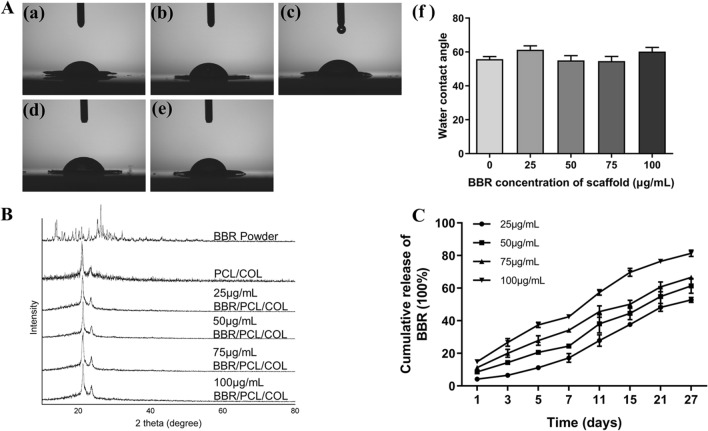
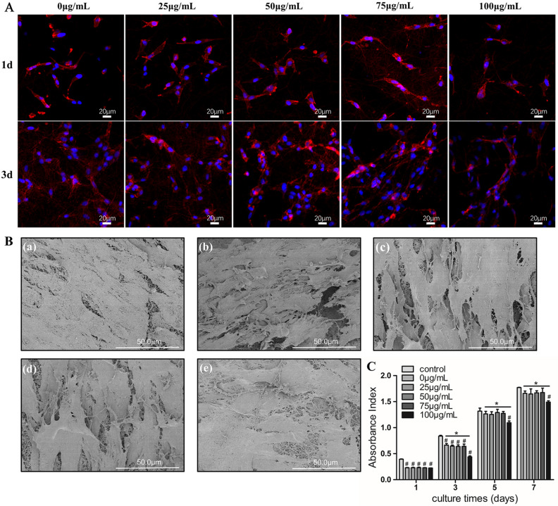
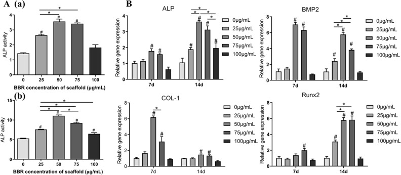
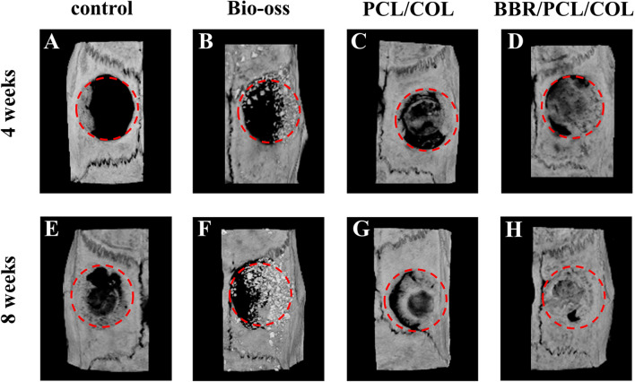
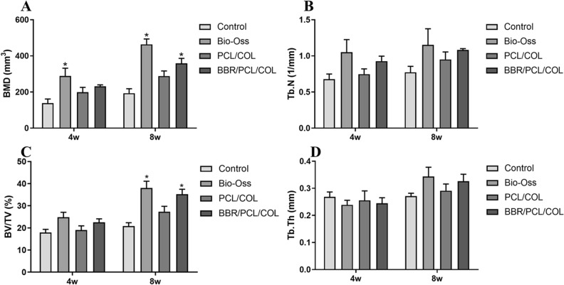
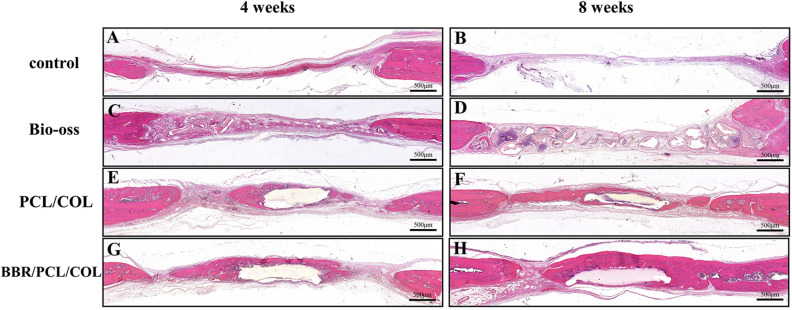
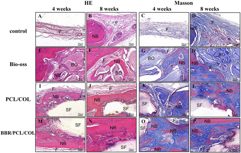
The repair of skeletal defects in maxillofacial region remains an intractable problem, the rising technology of bone tissue engineering provides a new strategy to solve it. Scaffolds, a crucial element of tissue engineering, must have favorable biocompatibility as well as osteoinductivity. In this study, we prepared berberine/polycaprolactone/collagen (BBR/PCL/COL) scaffolds with different concentrations of berberine (BBR) (25, 50, 75 and 100 μg/mL) through electrospinning. The influence of dosage on scaffold morphology, cell behavior and in vivo bone defect repair were systematically studied. The results indicated that scaffolds could release BBR stably for up to 27 days. Experiments in vitro showed that BBR/PCL/COL scaffolds had appropriate biocompatibility in the concentration of 25-75 μg/mL, and 50 and 75 μg/mL scaffolds could significantly promote osteogenic differentiation of dental pulp stem cells. Scaffold with 50 μg/mL BBR was implanted into the critical bone defect of rats to evaluate the ability of bone repair in vivo. It was found that BBR/PCL/COL scaffold performed more favorable than polycaprolactone/collagen (PCL/COL) scaffold. Overall, our study is the first to evaluate the capability of in vivo bone repair of BBR/PCL/COL electrospun scaffold. The results indicate that BBR/PCL/COL scaffold has prospective potential for tissue engineering applications in bone regeneration therapy.


Conflict of interest statement

The authors declare no competing interests.

Figures

Figure 1
Figure 1
Scaffolds morphology and elemental composition. (A) SEM images of PCL/COL scaffold (a) and BBR/PCL/COL (25, 50, 75 and 100 μg/mL) scaffolds (be). (B) The Cl elemental compositions of scaffolds.
Figure 2
Figure 2
Characterization of scaffolds. (A) Water contact angle analysis of scaffolds. (B) XRD spectra of PCL/COL scaffold, BBR/PCL/COL (25, 50, 75 and 100 μg/mL) scaffolds and BBR powder. (C) Cumulative release results of BBR from the BBR/PCL/COL (25, 50, 75 and 100 μg/mL) scaffolds.
Figure 3
Figure 3
The morphology and proliferation of DPSCs cultured on scaffolds. (A) DAPI and actinred immunocytochemical staining of DPSCs co-cultured on scaffolds after 1 day and 3 days. (B) The morphology of DPSCs cultured on scaffolds for 7 days. (a) PCL/COL scaffold; (b, c, d, e) BBR/PCL/COL (25, 50, 75 and 100 μg/mL) scaffolds. (C) Proliferation rate of DPSCs cultured on the different scaffolds at days 1, 3, 5, 7. “#” means the comparison between the control group and the experimental group, P < 0.05; “∗” means the comparison between experimental groups, P < 0.05.
Figure 4
Figure 4
Osteogenic differentiation of DPSCs cultured on scaffolds. (A) ALP activity of DPSCs co-cultured on scaffolds after 7 days (a) and 14 days (b). (B) Gene expressions of ALP, BMP2, COL-1, Runx2 in DPSCs co-cultured on the scaffolds after 7 days and 14 days. “#” means the comparison between the control group and the experimental group, P < 0.05; “∗” means the comparison between experimental groups, P < 0.05.
Figure 5
Figure 5
Images of micro-CT 3D reconstruction of rat calvaria defects at 4 weeks and 8 weeks after implantation.
Figure 6
Figure 6
Quantitative analysis of bone related parameters at 4 weeks and 8 weeks after implantation. “*” means the comparison between the control group and the experimental group, P < 0.05.
Figure 7
Figure 7
H&E staining of rat calvaria defects at 4 and 8 weeks after implantation.
Figure 8
Figure 8
H&E and Masson staining of defects at higher magnification. F, fibrous tissue; BO, Bio-oss; NB, new bone; SF, scaffold.

Similar articles

Cited by

References

    1. Huang RL, Kobayashi E, Liu K, Li Q. Bone graft prefabrication following the in vivo bioreactor principle. EBioMedicine. 2016;12:43–54. doi: 10.1016/j.ebiom.2016.09.016. - DOI - PMC - PubMed
    1. Salgado AJ, Coutinho OP, Reis RL. Bone tissue engineering: state of the art and future trends. Macromol. Biosci. 2004;4:743–765. doi: 10.1002/mabi.200400026. - DOI - PubMed
    1. Crane GM, Ishaug SL, Mikos AG. Bone tissue engineering. Nat. Med. 1995;1:1322–1324. doi: 10.1038/nm1295-1322. - DOI - PubMed
    1. Bouet G, Marchat D, Cruel M, Malaval L, Vico L. In vitro three-dimensional bone tissue models: From cells to controlled and dynamic environment. Tissue Eng. Part B Rev. 2015;21:133–156. doi: 10.1089/ten.TEB.2013.0682. - DOI - PubMed
    1. Hutmacher DW. Scaffolds in tissue engineering bone and cartilage. Biomaterials. 2000;21:2529–2543. doi: 10.1016/s0142-9612(00)00121-6. - DOI - PubMed

Publication types

MeSH terms

LinkOut - more resources