. 2021 Jan 13;11(1):927.
doi: 10.1038/s41598-020-80135-1.

Immune modulation resulting from MR-guided high intensity focused ultrasound in a model of murine breast cancer

Affiliations

Immune modulation resulting from MR-guided high intensity focused ultrasound in a model of murine breast cancer

Brett Z Fite et al. Sci Rep. .

Abstract

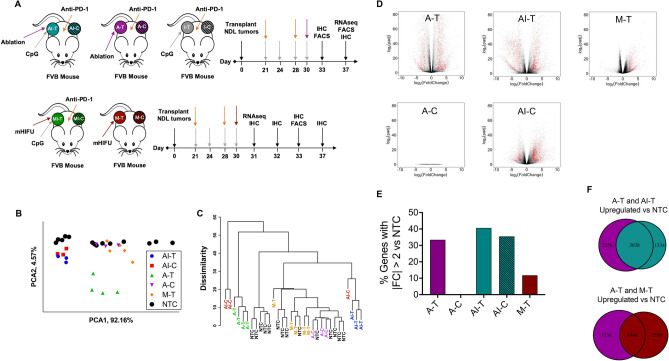
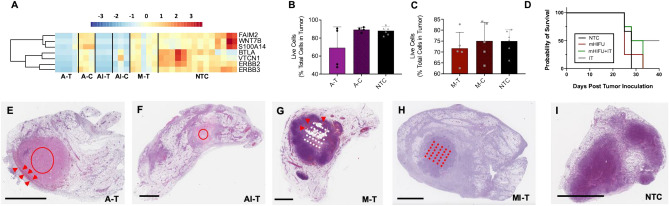
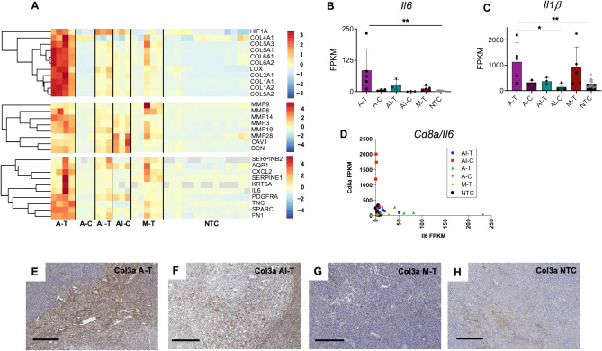
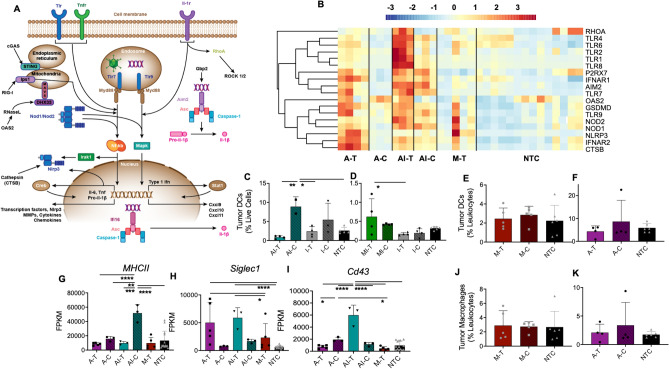
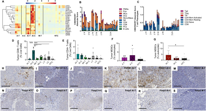
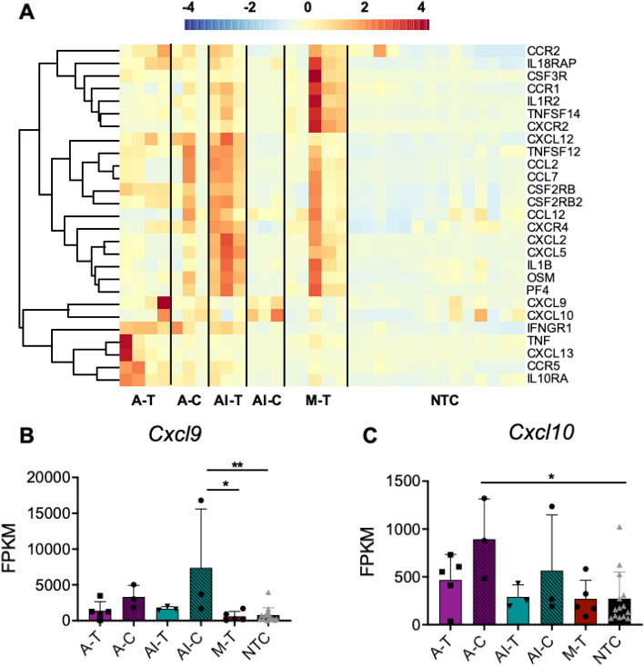
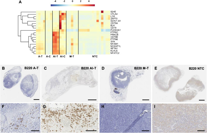
High intensity focused ultrasound (HIFU) rapidly and non-invasively destroys tumor tissue. Here, we sought to assess the immunomodulatory effects of MR-guided HIFU and its combination with the innate immune agonist CpG and checkpoint inhibitor anti-PD-1. Mice with multi-focal breast cancer underwent ablation with a parameter set designed to achieve mechanical disruption with minimal thermal dose or a protocol in which tumor temperature reached 65 °C. Mice received either HIFU alone or were primed with the toll-like receptor 9 agonist CpG and the checkpoint modulator anti-PD-1. Both mechanical HIFU and thermal ablation induced a potent inflammatory response with increased expression of Nlrp3, Jun, Mefv, Il6 and Il1β and alterations in macrophage polarization compared to control. Furthermore, HIFU upregulated multiple innate immune receptors and immune pathways, including Nod1, Nlrp3, Aim2, Ctsb, Tlr1/2/4/7/8/9, Oas2, and RhoA. The inflammatory response was largely sterile and consistent with wound-healing. Priming with CpG attenuated Il6 and Nlrp3 expression, further upregulated expression of Nod2, Oas2, RhoA, Pycard, Tlr1/2 and Il12, and enhanced T-cell number and activation while polarizing macrophages to an anti-tumor phenotype. The tumor-specific antigen, cytokines and cell debris liberated by HIFU enhance response to innate immune agonists.


Conflict of interest statement

The authors declare no competing interests.

Figures

Figure 1
Figure 1
HIFU reduces viable tumor and downregulates cancer and proliferation related genes and pathways. In all cases, “-T” indicates the directly-treated tumor and “-C” indicates the distant/contralateral tumor. (A) Schematic illustrating each treatment cohort and timing of treatments and endpoints for animals treated with (top) thermal ablation and (bottom) mechanical HIFU. (B) Principal component analysis biplot of the first two components of treatment cohorts. (C) Hierarchical clustering dendrogram of the entire transcriptome for each individual tumor within each treatment cohort. (D) Volcano plots of each treatment cohort plotting log2 fold change versus negative log2 adjusted p-value of each gene’s expression compared to control, plotted only for genes whose expression is significantly changed (p < 0.05). Red points indicate genes where the absolute value of the log2 fold change is greater than 2. (E) Bar plot summarizing the percentage of genes with expression |FC|> 2 out of all significantly altered genes for each cohort compared to control. (F) Venn diagrams illustrating overlap between thermal ablation and thermal ablation with immunotherapy and between thermal ablation and mechanical HIFU of upregulated genes (compared to control). Volcano plots, Venn diagrams, and the hierarchical clustering dendrogram were created in R Studio v1.2.5001 (https://rstudio.com).
Figure 2
Figure 2
HIFU reduces viable tumor and downregulates cancer and proliferation related genes and pathways. (A) Heatmap visualization of Z-scores of cancer and proliferation-related genes for each treatment cohort. Live cells as measured with flow cytometry of the (B) thermal ablation and (C) mechanical ablation cohorts. (D) Survival proportions of control animals compared to animals that were treated with mechanical HIFU alone, immunotherapy alone, or the combination of mechanical HIFU and immunotherapy. H&E sections of (E) a thermally-ablated tumor (arrows indicate rim of viable tumor cells), (F) a tumor that received both thermal ablation and immunotherapy, (G) a tumor treated with mechanical HIFU (arrows indicate hemorrhage), (H) a tumor that received both mechanical HIFU and immunotherapy, and (I) an untreated control tumor. Scale bar represents 2 mm in all cases. Time points for RNA-seq are: 1 week post ablation for all cohorts that received thermal ablation, and 24 h post HIFU for cohorts that received mechanical HIFU. For flow cytometry studies, time points are 72 h for cohorts that received mechanical HIFU and 1 week for cohorts that received thermal ablation alone. Time points for IHC are 72 h post ablation for thermal ablation alone, 24 h for mechanical HIFU alone, and 1 week for both thermal and mechanical ablation plus immunotherapy. Red and white overlays on histological sections indicate the insonation pattern performed (either circular continuous wave for thermal ablation or a grid pattern for mechanical HIFU). The heatmap was created in R Studio v1.2.5001 (https://rstudio.com).
Figure 3
Figure 3
HIFU induces strong inflammatory response. (A) Z-scores of ECM remodeling related (top two panes) and wound healing related (bottom pane) genes for each treatment cohort. FPKM values for each cohort for (B) the inflammatory cytokine IL6, (C) an upstream regulator of inflammation IL1b. Scatter plot of the FPKM value ratios of (D) Cd8, versus Il6 for each cohort. Col3a IHC stained sections of (E) a thermally-ablated tumor, (F) a tumor that received both thermal ablation and immunotherapy, (G) a tumor treated with mechanical HIFU, and (H) an untreated control tumor. Scale bar represents 200 µm in all cases. Time points are 1-week post ablation for all cohorts that received thermal ablation, and 24 h post HIFU for cohorts that received mechanical HIFU. Data are expressed as mean ± SD. *p < 0.05, **p < 0.01 (ordinary one-way ANOVA with Tukey correction for multiple hypotheses). The heatmap was created in R Studio v1.2.5001 (https://rstudio.com).
Figure 4
Figure 4
HIFU stimulates innate immune sensors and generates an innate response when combined with immunotherapy. (A) Schematic of innate immune receptors and signaling. (B) Heatmap visualization of Z-scores of innate immune sensors. Dendritic cells quantified with flow cytometry of the (C) thermal ablation pre-treated with immunotherapy, (D) mechanical HIFU pre-treated with immunotherapy, (E) mechanical HIFU, and (F) thermal ablation cohorts. FPKM values for each cohort for (G) MHCII, (H) Siglec1, and (I) Cd43. Macrophages as measured with flow cytometry of the (J) mechanical HIFU and (K) thermal ablation cohorts. Time points for RNA-seq are 1-week post ablation for all cohorts that received thermal ablation, and 24 h post HIFU for cohorts that received mechanical HIFU. For flow cytometry studies, time points are 72 h for cohorts that received mechanical HIFU or immunotherapy in addition to thermal ablation and 1 week for cohorts that received thermal ablation alone. Data are expressed as mean ± SD. *p < 0.05, **p < 0.01, ***p < 0.001, ****p < 0.0001 (ordinary one-way ANOVA with Tukey correction for multiple hypotheses). The heatmap was created in R Studio v1.2.5001 (https://rstudio.com).
Figure 5
Figure 5
Thermal ablation stimulates a weak adaptive response that is augmented by TLR ligation and checkpoint modulation. (A) Z-scores of adaptive immune and T-cell response related genes for each treatment cohort. (B) CIBERSORTx-imputed absolute immune cell content. (C) CIBERSORTx-imputed T-cell phenotype distribution. Flow cytometry quantitation of (D) CD8+ and (E) CD4+ T-cells in tumors treated with a combined immunotherapy-HIFU protocol. Myeloid derived suppressor cells (MDSCs) as quantified with flow cytometry of the (F) thermal ablation and (G) mechanical ablation cohorts. Pdcd1 IHC stained sections of (H) an untreated control tumor, (I) a thermally-ablated tumor, (J) a distant tumor in an animal that received thermal ablation, (K) a tumor that received both thermal ablation and immunotherapy, (L) a distant tumor in an animal that received both thermal ablation and immunotherapy, and (M) a tumor treated with mechanical HIFU. FoxP3 IHC stained sections of (N) an untreated control tumor, (O) a thermally-ablated tumor, (P) a distant tumor in an animal that received thermal ablation, (Q) a tumor that received both thermal ablation and immunotherapy, (R) a distant tumor in an animal that received both thermal ablation and immunotherapy, and (S) a tumor treated with mechanical HIFU. Scale bar represents 100 µm in all cases. Time points for RNA-seq and IHC data are: 1-week post ablation for all cohorts that received thermal ablation, and 24 h post HIFU for cohorts that received mechanical HIFU. For flow cytometry studies, time points are 72 h for cohorts that received mechanical HIFU or immunotherapy in addition to thermal ablation and 1 week for cohorts that received thermal ablation alone. Data are expressed as mean ± SD. *p < 0.05, **p < 0.01, ****p < 0.0001 (ordinary one-way ANOVA with Tukey correction for multiple hypotheses). The heatmap was created in R Studio v1.2.5001 (https://rstudio.com).
Figure 6
Figure 6
HIFU upregulates inflammatory cytokines and leukocyte signaling chemokines. (A) Heatmap visualization of Z-scores of leukocyte chemokines and cytokines for each treatment cohort. FPKM values for each cohort for (B) Cxcl9 and (C) Cxcl10. Data are expressed as mean ± SD. *p < 0.05, **p < 0.01 (ordinary one-way ANOVA with Tukey correction for multiple hypotheses). Time points are: 1-week post ablation for all cohorts that received thermal ablation, and 24 h post HIFU for cohorts that received mechanical HIFU. The heatmap was created in R Studio v1.2.5001 (https://rstudio.com).
Figure 7
Figure 7
Thermal ablation recruits B220+ cells and upregulates pathways consistent with plasma-resident immune cell signaling and activation. (A) Z-scores of plasma immune cell-related genes for each treatment cohort. B220 stains of (B) thermally-ablated tumor, (C) tumor that received both thermal ablation and immunotherapy, (D) tumor treated with mechanical HIFU, and (E) untreated control tumor. Magnified views of the same B220-stained sections for (F) thermally-ablated tumor, (G) tumor that received both thermal ablation and immunotherapy, (H) tumor treated with mechanical HIFU, and (I) an untreated control tumor. Scale bar represents 2 mm in (B–E) and 100 µm in (FI). Time points are: 1-week post ablation for all cohorts that received thermal ablation, and 24 h post HIFU for cohorts that received mechanical HIFU. The heatmap was created in R Studio v1.2.5001 (https://rstudio.com).

Similar articles

Cited by

References

    1. Ning Z, et al. HIFU is safe, effective, and feasible in pancreatic cancer patients: A monocentric retrospective study among 523 patients. Onco Targets Ther. 2019;12:1021–1029. doi: 10.2147/OTT.S185424. - DOI - PMC - PubMed
    1. Strunk HM, et al. Clinical use of high-intensity focused ultrasound (HIFU) for tumor and pain reduction in advanced pancreatic cancer. Rofo. 2016;188:662–670. doi: 10.1055/s-0042-105517. - DOI - PubMed
    1. Khokhlova TD, Hwang JH. HIFU for palliative treatment of pancreatic cancer. Adv Exp. Med. Biol. 2016;880:83–95. doi: 10.1007/978-3-319-22536-4_5. - DOI - PubMed
    1. de Senneville BD, Moonen C, Ries M. MRI-guided HIFU methods for the ablation of liver and renal cancers. Adv. Exp. Med. Biol. 2016;880:43–63. doi: 10.1007/978-3-319-22536-4_3. - DOI - PubMed
    1. Fite BZ, et al. Magnetic resonance thermometry at 7T for real-time monitoring and correction of ultrasound induced mild hyperthermia. PLoS ONE. 2012;7:e35509. doi: 10.1371/journal.pone.0035509. - DOI - PMC - PubMed

Publication types

MeSH terms