. 2021 Jan 13;11(1):1034.
doi: 10.1038/s41598-020-80524-6.

Tethering-induced destabilization and ATP-binding for tandem RRM domains of ALS-causing TDP-43 and hnRNPA1

Affiliations

Tethering-induced destabilization and ATP-binding for tandem RRM domains of ALS-causing TDP-43 and hnRNPA1

Mei Dang et al. Sci Rep. .

Abstract

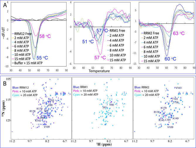
TDP-43 and hnRNPA1 contain tandemly-tethered RNA-recognition-motif (RRM) domains, which not only functionally bind an array of nucleic acids, but also participate in aggregation/fibrillation, a pathological hallmark of various human diseases including amyotrophic lateral sclerosis (ALS), frontotemporal dementia (FTD), alzheimer's disease (AD) and Multisystem proteinopathy (MSP). Here, by DSF, NMR and MD simulations we systematically characterized stability, ATP-binding and conformational dynamics of TDP-43 and hnRNPA1 RRM domains in both tethered and isolated forms. The results reveal three key findings: (1) upon tethering TDP-43 RRM domains become dramatically coupled and destabilized with Tm reduced to only 49 °C. (2) ATP specifically binds TDP-43 and hnRNPA1 RRM domains, in which ATP occupies the similar pockets within the conserved nucleic-acid-binding surfaces, with the affinity slightly higher to the tethered than isolated forms. (3) MD simulations indicate that the tethered RRM domains of TDP-43 and hnRNPA1 have higher conformational dynamics than the isolated forms. Two RRM domains become coupled as shown by NMR characterization and analysis of inter-domain correlation motions. The study explains the long-standing puzzle that the tethered TDP-43 RRM1-RRM2 is particularly prone to aggregation/fibrillation, and underscores the general role of ATP in inhibiting aggregation/fibrillation of RRM-containing proteins. The results also rationalize the observation that the risk of aggregation-causing diseases increases with aging.


Conflict of interest statement

The authors declare no competing interests.

Figures

Figure 1
Figure 1
Dissection-induced perturbation of TDP-43 RRM domains. (A) 414-residue TDP-43 contains: N-terminal domain (NTD) over residues 1–80; two tandemly-tethered RNA recognition motifs (RRM1 and RRM2) over residues 105–261, and C-terminal prion-like domain over residues 274–414. (B) NMR structure (PDB ID of 4BS2) of the TDP-43 RRM1 (light blue) and RRM2 (pink) in which the residues are displayed in sphere for those with HSQC peaks disappeared (yellow), significantly shifted in RRM1 (blue) or in RRM2 (light pink) upon dissection. The image was prepared by PyMol2.4 (https://pymol.org). (C) Superimposition of 1H-15 N NMR HSQC spectra of the 15 N-labeled tethered RRM1–RRM2 domains (blue), the isolated RRM1 (cyan) and RRM2 (pink) proteins at 50 μM in 10 mM sodium phosphate buffer (pH 6.8) containing 150 mM NaCl and 10 mM DTT. (D) Residue-specific chemical shift difference (CSD) of the RRM1 and RRM2 domains between the tethered and isolated forms. Significantly shifted residues are labeled and displayed as spheres in (B), which are defined as those with the CSD values > 0.25 (average value + one standard deviation) (pink line).
Figure 2
Figure 2
Thermal stability and ATP binding of the isolated TDP-43 RRM2. (A) DSF melting curves of thermal unfolding of the isolated RRM2 domain in the presence of ATP at different concentrations. (B) 1H-15 N NMR HSQC spectra of the 15 N-labeled RRM2 domain at 50 μM (blue) in 10 mM sodium phosphate buffer (pH 6.8) containing 150 mM NaCl and 10 mM DTT in the absence (blue), and in the presence of ATP at 10 (pink) and 20 (cyan) mM. Traces of two representative HSQC peaks, which have significant shifts but are not severely overlapped with other peaks. For clarity, only peaks at five ATP concentrations are shown: in the free state (blue); in the presence of ATP at 6 mM (pink); 10 mM (cyan); 14 mM (black); and 20 mM (red). (C) Residue-specific chemical shift difference (CSD) of the isolated RRM2 in the presence of ATP at 10 mM (blue) and 20 mM (pink). Significantly shifted residues are labeled, which are defined as those with the CSD values at 20 mM ATP > 0.1 (average value + one standard deviation) (cyan line). Fitting of the residue-specific dissociation constant (Kd): experimental (dots) and fitted (lines) values for the chemical shift differences induced by addition of ATP at 0.5, 1.0, 2.0, 4.0, 6.0, 8.0, 10.0, 12.0, 14.0, 16.0, 18.0 and 20.0 mM.
Figure 3
Figure 3
Dissection-induced perturbation of hnRNPA1 RRM domains. (A) 320-residue hnRNPA1 contains: two RNA recognition motifs (RRM1 and RRM2) over residues 1–179, and C-terminal prion-like domain over residues 180–320. (B) NMR structure (PDB ID of 2LYV) of the hnRNPA1 RRM1 (light blue) and RRM2 (pink) in which the residues are displayed in spheres for those with HSQC peaks significantly shifted in RRM1 (blue) or in RRM2 (light pink) upon dissection. The image was prepared by PyMol2.4 (https://pymol.org). (C) Superimposition of 1H-15 N NMR HSQC spectra of the 15 N-labeled tethered RRM1–RRM2 domains (blue), the isolated RRM1 (cyan) and RRM2 (pink) domains at 50 μM in 10 mM sodium phosphate buffer (pH 6.8) containing 150 mM NaCl and 10 mM DTT. (D) Residue-specific chemical shift difference (CSD) of the RRM1 and RRM2 domains between the tethered and isolated forms. Significantly shifted residues are labeled and displayed as spheres, which are defined as those with the CSD values > 0.09 (average value + one standard deviation) (pink line).
Figure 4
Figure 4
Thermal stability and ATP binding of the hnRNPA1 RRM domains. (A) DSF melting curves of thermal unfolding of the tethered RRM1–RRM2, as well as isolated RRM1 and RRM2 domains in the presence of ATP at different concentrations. (B) 1H-15 N NMR HSQC spectra of the 15 N-labeled tethered RRM1–RRM2, as well as isolated RRM1 and RRM2 domains at 50 μM (blue) in 10 mM sodium phosphate buffer (pH 6.8) containing 150 mM NaCl and 10 mM DTT in the absence (blue), and in the presence of ATP at 10 (pink) and 20 (cyan) mM.
Figure 5
Figure 5
ATP binding of the tethered RRM1–RRM2, as well as isolated RRM2 of hnRNPA1. (A) Residue-specific chemical shift difference (CSD) of the tethered hnRNPA1 RRM1–RRM2 in the presence of ATP at 10 mM (blue) and 20 mM (pink). Significantly shifted residues are labeled, which are defined as those with the CSD values at 20 mM ATP > 0.1 (average value + one standard deviation) (cyan line). (B) Residue-specific chemical shift difference (CSD) of the isolated RRM2 in the presence of ATP at 10 mM (blue) and 20 mM (pink). Significantly shifted residues are labeled, which are defined as those with the CSD values at 20 mM ATP > 0.125 (average value + one standard deviation) (cyan line). The shifted residues identical to those in the tethered form are labeled in pink while the additionally shifted residues are in black. (C) The lowest energy docking model of the ATP-RRM2 complex. The structure of hnRNPA1 RRM2 is displayed in ribbon, while ATP is in sticks. The nine residues with significant CSD values are displayed in spheres and labeled. (D) Superimposition of three complexes of ATP: with hnRNPA1 RRM2 (pink), TDP-43 RRM1 (yellow) and TDP-43 RRM2 (cyan). (E) The ATP-RRM2 complex of hnRNPA1 with the RRM2 structure displayed in the electrostatic potential surface and ATP in sticks. (F) The ATP-RRM complex of hnRNPA1 showing three hydrogen bonds (in blue dotted lines) between ATP and RRM2 atoms. (G) Superimposition of the hnRNPA1 RRM1 and ATP-RRM2 complex with the three significantly shifted RRM1 residue (Arg88, Val90 and Ser91) displayed in spheres. The image was prepared by PyMol2.4 (https://pymol.org).
Figure 6
Figure 6
Overall dynamic behaviors of the TDP-43 RRM domains. (A) Average root-mean-square deviations (RMSD) of the Cα atoms over three independent 50-ns MD simulations for the tethered RRM1–RRM2 (black), as well as isolated RRM1 (blue) and RRM2 (pink) (I). For the RRM1 in the tethered form (black) and isolated form (blue) with the unstructured N- and C-termini excluded for calculation (II); as well as for the RRM2 in the tethered form (black) and isolated form (pink) with the unstructured N- and C-termini excluded for calculation (III). (B) Structure snapshots in the first MD simulation with one structure for each 5 ns. The image was prepared by PyMol2.4 (https://pymol.org).
Figure 7
Figure 7
Residue-specific dynamic behaviors of the TDP-43 RRM domains. (A) Averaged root-mean-square fluctuations (RMSF) of the Cα atoms computed over three independent MD simulations for the tethered RRM1–RRM2 (black), as well as isolated RRM1 (blue) and RRM2 (pink). (B) The difference of the average RMSF between the tethered RRM1–RRM2, and isolated RRM1/RRM2. The residues are labeled for those with significant difference of RMSF (> 1.0; average + one STD). (C) Structure of the TDP-43 RRM1–RRM2 with the residues displayed in spheres which show significant difference of RMSF between the tethered and isolated forms. The image was prepared by PyMol2.4 (https://pymol.org). (D) Mutual information matrix calculated from three parallel MD simulation data of the tethered TDP-43 RRM1–RRM2 by MutInf, with residues having significant inter-domain correlation motions highlighted by yellow boxes. The RRM1 sequence is indicated by blue box and RRM2 sequence in pink box.
Figure 8
Figure 8
Overall dynamic behaviors of the hnRNPA1 RRM domains. (A) Average root-mean-square deviations (RMSD) of the Cα atoms over three independent 50-ns MD simulations for the tethered RRM1–RRM2 (black), as well as isolated RRM1 (blue) and RRM2 (pink) (I). for the RRM1 in the tethered form (black) and isolated form (blue) with the unstructured N- and C-termini excluded for calculation (II); as well as for the RRM2 in the tethered form (black) and isolated form (pink) with the unstructured N- and C-termini excluded for calculation (III). (B) Structure snapshots in the first MD simulation with one structure for each 5 ns. The image was prepared by PyMol2.4 (https://pymol.org).
Figure 9
Figure 9
Residue-specific dynamic behaviors of the hnRNPA1 RRM domains. (A) Averaged root-mean-square fluctuations (RMSF) of the Cα atoms computed over three independent MD simulations for the tethered RRM1–RRM2 (black), as well as isolated RRM1 (blue) and RRM2 (pink). (B) The difference of the average RMSF between the tethered RRM1–RRM2, and isolated RRM1/RRM2. The residues are labeled for those with significant difference of RMSF (> 2.0; average + one STD). (C) Structure of the hnRNPA1 RRM1–RRM2 with the residues displayed in spheres which show significant difference of RMSF between the tethered and isolated states. The image was prepared by PyMol2.4 (https://pymol.org). (D) Mutual information matrix calculated from three parallel MD simulation data of the tethered hnRNPA1 RRM1–RRM2 by MutInf, with residues having significant inter-domain correlation motions highlighted by yellow box. The RRM1 sequence is indicated by blue box and RRM2 sequence in pink box.

Similar articles

Cited by

References

    1. Neumann M, et al. Ubiquitinated TDP-43 in frontotemporal lobar degeneration and amyotrophic lateral sclerosis. Science. 2006;314:130–133. doi: 10.1126/science.1134108. - DOI - PubMed
    1. Ling SC, Polymenidou M, Cleveland DW. Converging mechanisms in ALS and FTD: Disrupted RNA and protein homeostasis. Neuron. 2013;79:416–438. doi: 10.1016/j.neuron.2013.07.033. - DOI - PMC - PubMed
    1. Harrison AF, Shorter J. RNA-binding proteins With Prion-Like domains in health and disease. Biochem J. 2017;474:1417–1438. doi: 10.1042/BCJ20160499. - DOI - PMC - PubMed
    1. Geuens T, Bouhy D, Timmerman V. The hnRNP family: Insights into their role in health and disease. Hum. Genet. 2016;135:851–867. doi: 10.1007/s00439-016-1683-5. - DOI - PMC - PubMed
    1. Cléry AM, Blatter M, Allain FH. RNA recognition motifs: Boring? Not. Quite Curr. Opin. Struct. Biol. 2008;18:290–298. doi: 10.1016/j.sbi.2008.04.002. - DOI - PubMed

Publication types

MeSH terms

Substances