. 2021 Jan 13;11(1):949.
doi: 10.1038/s41598-020-80294-1.

Exosite inhibition of ADAMTS-5 by a glycoconjugated arylsulfonamide

Affiliations

Exosite inhibition of ADAMTS-5 by a glycoconjugated arylsulfonamide

Salvatore Santamaria et al. Sci Rep. .

Abstract

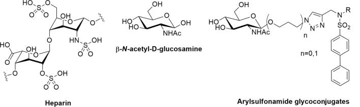
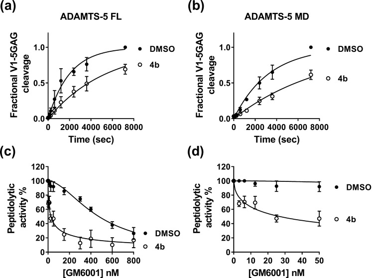
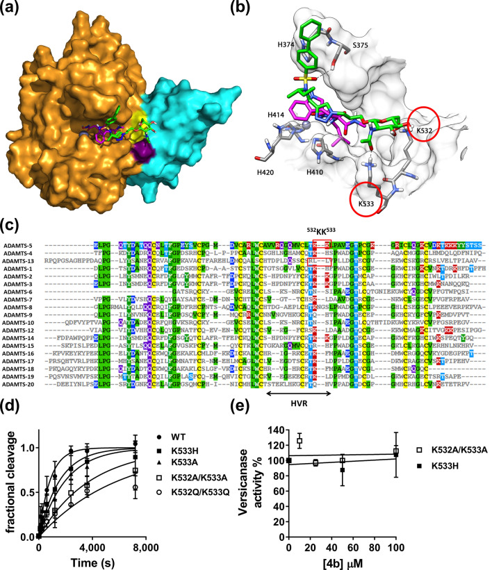
ADAMTS-5 is a major protease involved in the turnover of proteoglycans such as aggrecan and versican. Dysregulated aggrecanase activity of ADAMTS-5 has been directly linked to the etiology of osteoarthritis (OA). For this reason, ADAMTS-5 is a pharmaceutical target for the treatment of OA. ADAMTS-5 shares high structural and functional similarities with ADAMTS-4, which makes the design of selective inhibitors particularly challenging. Here we exploited the ADAMTS-5 binding capacity of β-N-acetyl-D-glucosamine to design a new class of sugar-based arylsulfonamides. Our most promising compound, 4b, is a non-zinc binding ADAMTS-5 inhibitor which showed high selectivity over ADAMTS-4. Docking calculations combined with molecular dynamics simulations demonstrated that 4b is a cross-domain inhibitor that targets the interface of the metalloproteinase and disintegrin-like domains. Furthermore, the interaction between 4b and the ADAMTS-5 Dis domain is mediated by hydrogen bonds between the sugar moiety and two lysine residues (K532 and K533). Targeted mutagenesis of these two residues confirmed their importance both for versicanase activity and inhibitor binding. This positively-charged cluster of ADAMTS-5 represents a previously unknown substrate-binding site (exosite) which is critical for substrate recognition and can therefore be targeted for the development of selective ADAMTS-5 inhibitors.


Conflict of interest statement

The authors declare no competing interests.

Figures

Figure 1
Figure 1
Comparison between heparin and the glycoconjugates analyzed in this study. R represents either a ZBG or an aliphatic moiety.
Figure 2
Figure 2
Inhibitory activity of compound 4b. (A) Inhibition of versicanase activity. ADAMTS-4 (5.5 nM) and -5 (0.4 nM) were incubated either with compound 4b or DMSO for 2 h at 37 °C before addition of V1-5GAG (50 nM). At each time point, reactions were stopped by addition of EDTA and ADAMTS-generated versican fragments (versikine) quantified by sandwich ELISA. The relative versicanase activity is presented and 100% activity corresponds to that in the presence of DMSO alone. (B) Inhibition of ADAMTS-5 aggrecanase activity. Compounds (Cpd) 4b, 5b and 6 were incubated with ADAMTS-5 (1 nM) for 2 h at 37 °C before addition of aggrecan (20 μg). Following SDS-PAGE and immunoblot, fragments cleaved at the Glu392↓Ala393 bond were detected by a monoclonal neoepitope antibody recognizing the new C-terminal fragment (anti-ARGSV) and analyzed by densitometric analysis. Data are presented as mean ± SEM (n = 4). *p < 0.05 and ***p < 0.001, compared to DMSO controls; ##p < 0.01, compared to the same concentration of compound 6 (Mann–Whitney test).
Figure 3
Figure 3
Compound 4b binds to the ADAMTS-5 Mp/Dis domains in the presence of GM6001. (A, B) Compound 4b inhibits versicanase activity of both full-length (FL) ADAMTS-5 and its variant containing only the Mp/Dis domains (ADAMTS-5 MD). Compound 4b (100 μM) or DMSO was incubated either with ADAMTS-5 FL (0.2 nM, A) or ADAMTS-5 MD (26 nM, B) for 2 h at 37 °C before addition of V1-5GAG (50 nM). At each time point, reactions were stopped by addition of EDTA and versikine fragments quantified by sandwich ELISA as reported in the Methods section. In the DMSO control, complete proteolysis was achieved after 7200 s. (C, D) Inhibition of ADAMTS-5 peptidolytic activity by the active-site inhibitor GM6001 in the presence of compound 4b. ADAMTS-5 MDTCS was incubated with active-site inhibitor GM6001 (0–800 nM) either in the presence of DMSO or compound 4b (10 μM) for 2 h at 37 °C before addition of QF peptide. The synergistic effect exerted by compound 4b is more evident at low concentrations of GM6001 (panel D). The relative peptidolytic activity is presented and 100% activity corresponds to that in the presence of DMSO alone. Data are presented as mean ± SEM (n = 3).
Figure 4
Figure 4
Compound 4b binds to an exosite in the ADAMTS-5 Dis domain. (A, B) Minimized average structure of compound 4b bound to the ADAMTS-5-GM6001 complex, derived from the last 100 ns of MD simulation. (A) Protein surface. 4b is shown in green, GM6001 is in pink, ADAMTS-5 Mp domain in gold, the Dis domain in light blue, the active site zinc as a grey sphere. Exosite residues K532 and K533 are colored in yellow and purple, respectively. (B) Protein residues directly interacting with compound 4b. H-bonds are represented as black dashed lines. (C) Amino acid sequence alignment of the Dis domain of all ADAMTS family members. Known exosites are boxed and the poorly conserved region (HVR, hypervariable region) is indicated. Conserved residues are indicated by the same color code. (D) Time course experiments for cleavage of 50 nM V1-5GAG by ADAMTS-5 MDTCS variants. Enzyme (0.4 nM) was incubated with 50 nM substrate. At the indicated time points, an aliquot was taken, stopped with EDTA and cleavage products measured by sandwich ELISA as described in the Methods section. The solid lines represent a nonlinear regression fit of the data. The data are presented as average ± SEM; n = 3–4. (E) Inhibition of ADAMTS-5 MDTCS variants by compound 4b. ADAMTS-5 K533H and K532A/K533A (0.82 nM) were incubated either with compound 4b or DMSO for 2 h at 37 °C before addition of V1-5GAG (50 nM). At each time point, reactions were stopped by addition of EDTA and cleavage fragments quantified by sandwich ELISA. The relative versicanase activity is presented and 100% activity corresponds to that in the presence of DMSO alone. The data are presented as average ± SEM; n = 3.

References

    1. Kiani C, Chen L, Wu YJ, Yee AJ, Yang BB. Structure and function of aggrecan. Cell Res. 2002;12:19–32. doi: 10.1038/sj.cr.7290106. - DOI - PubMed
    1. Wight TN. A role for proteoglycans in vascular disease. Matrix Biol. 2018;71–72:396–420. doi: 10.1016/j.matbio.2018.02.019. - DOI - PMC - PubMed
    1. Heinegård D, Saxne T. The role of the cartilage matrix in osteoarthritis. Nat. Rev. Rheumatol. 2011;7:50–56. doi: 10.1038/nrrheum.2010.198. - DOI - PubMed
    1. Fushimi K, Troeberg L, Nakamura H, Lim NH, Nagase H. Functional differences of the catalytic and non-catalytic domains in human ADAMTS-4 and ADAMTS-5 in aggrecanolytic activity. J. Biol. Chem. 2008;283:6706–6716. doi: 10.1074/jbc.M708647200. - DOI - PubMed
    1. Santamaria S, et al. Exosites in hypervariable loops of ADAMTS spacer domains control substrate recognition and proteolysis. Sci. Rep. 2019;9:10914. doi: 10.1038/s41598-019-47494-w. - DOI - PMC - PubMed

Publication types

LinkOut - more resources