. 2021 Jan 13;11(1):1141.
doi: 10.1038/s41598-020-80881-2.

Comprehensive analysis of metastatic gastric cancer tumour cells using single-cell RNA-seq

Affiliations

Comprehensive analysis of metastatic gastric cancer tumour cells using single-cell RNA-seq

Bin Wang et al. Sci Rep. .

Abstract

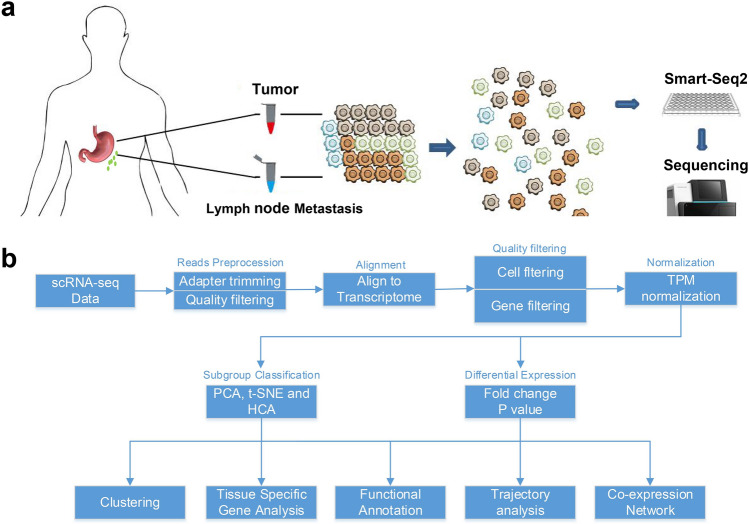
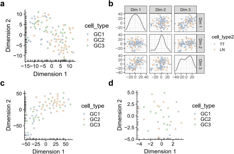
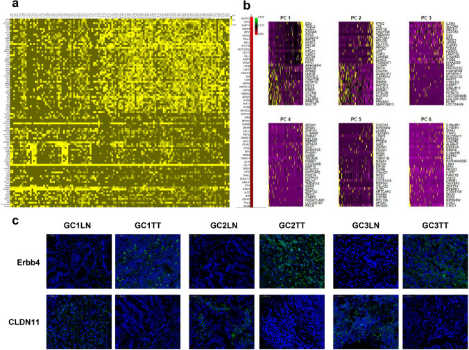
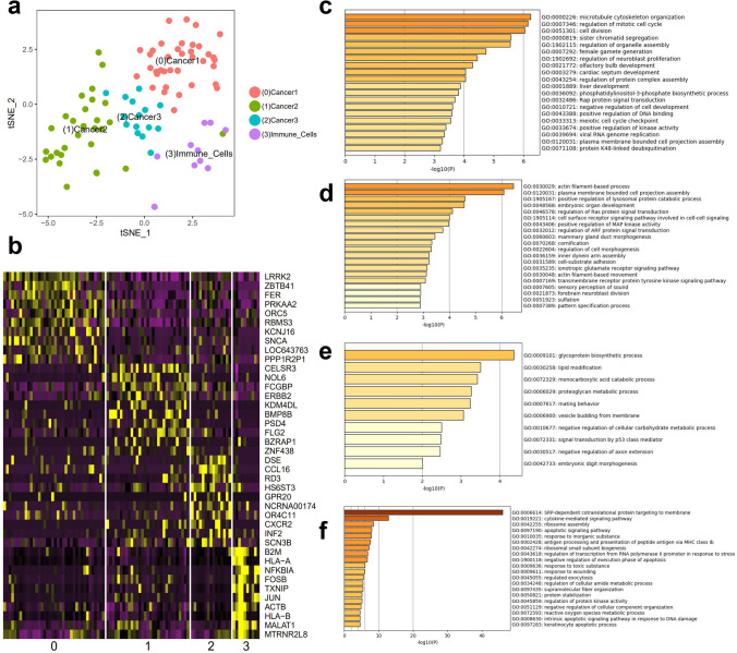
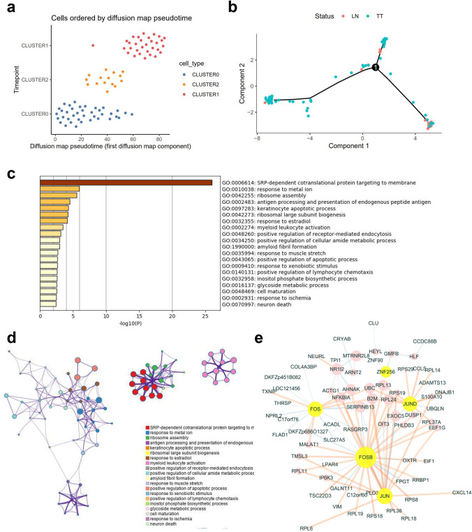
Gastric cancer (GC) is a leading cause of cancer-induced mortality, with poor prognosis with metastasis. The mechanism of gastric carcinoma lymph node metastasis remains unknown due to traditional bulk-leveled approaches masking the roles of subpopulations. To answer questions concerning metastasis from the gastric carcinoma intratumoural perspective, we performed single-cell level analysis on three gastric cancer patients with primary cancer and paired metastatic lymph node cancer tissues using single-cell RNA-seq (scRNA-seq). The results showed distinct carcinoma profiles from each patient, and diverse microenvironmental subsets were shared across different patients. Clustering data showed significant intratumoural heterogeneity. The results also revealed a subgroup of cells bridging the metastatic group and primary group, implying the transition state of cancer during the metastatic process. In the present study, we obtained a more comprehensive picture of gastric cancer lymph node metastasis, and we discovered some GC lymph node metastasis marker genes (ERBB2, CLDN11 and CDK12), as well as potential gastric cancer evolution-driving genes (FOS and JUN), which provide a basis for the treatment of GC.


Conflict of interest statement

The authors declare no competing interests.

Figures

Figure 1
Figure 1
(a) Overview of the study design and sampling protocol. (b) Analysis pipeline in the current single-cell RNA-seq study.
Figure 2
Figure 2
(a) T-SNE was plotted to present the distribution of the single cells from three patients in all primary tumour tissues. (b) Unsupervised T-SNE showing the separation of carcinoma cell groups. (c) In terms of gastric cancer tumour tissues, removal of noncarcinoma cells reveals intrinsic patient-specific tumour cell heterogeneity. (d) More randomly dispersed dots are shown in the metastatic tumour cells.
Figure 3
Figure 3
Intratumoural heterogeneity analysis. Correlation analysis of single cells revealed heterogeneity within tumours across three patients. Robust bulk stemness, immune, stromal, and tumour scoring assessment between tumour and paratumour single cells.
Figure 4
Figure 4
Tissue-specific markers. (a) Population-wide comparison between TT and LN single cells. Tissue-specific markers were calculated, and a heatmap was plotted using the top 100 highly expressed features based on previously defined clusters. NOTCH2, NOTCH2NL, KIF5B, and ERBB4 are highly expressed in primary cancer, while CDK12, ERBB2, and CLDN11 are overexpressed in metastatic cancer. Functional annotation revealed microtubule movement, and notch-based signalling was activated in the primary cells, indicating its metastatic propensity. (b) Decomposition of six main principal components (PCs) in the datasets. (c) IF of ERBB4 and CLDN11 in TT and LN.
Figure 5
Figure 5
Seurat marker analysis revealed four main clusters in the overall single cells. Twelve significant principal components were extracted to identify four main clusters in the tumour tissues. (a) Four main clusters in the tumour tissues using TSNE. (b) Heatmap indicating markers highly expressed in each cluster. (cf) Functional annotations of each cluster are shown based on Seurat-calculated markers.
Figure 6
Figure 6
Gastric-derived cell evolutionary trajectory. (a) The pseudotime trajectory of GC clusters revealed a distinct pattern of postulated evolution state from Cluster 0 > 2 > 1. (b) The evolutionary trajectory of TT and LN cells. (c,d) Evolution trajectory-based functional annotation (Top1000 gene). (e) Regulatory co-network of kernel genes in evolution regulation and a transcription factor-driven regulatory network.

References

    1. McGuire, S. World Cancer Report Geneva, Switzerland: World Health Organization, International Agency for Research on Cancer, WHO Press, 2015. Adv. Nutr. 2014;7(418–419):2016. doi: 10.3945/an.116.012211. - DOI - PMC - PubMed
    1. Aurello P, et al. Classification of lymph node metastases from gastric cancer: Comparison between N-site and N-number systems. Our experience and review of the literature. Am. Surgeon. 2007;73:359–366. doi: 10.1177/000313480707300410. - DOI - PubMed
    1. Grimes JA, et al. Agreement between cytology and histopathology for regional lymph node metastasis in dogs with melanocytic neoplasms. Vet. Pathol. 2017;54:579–587. doi: 10.1177/0300985817698209. - DOI - PubMed
    1. Ehteshami Bejnordi B, et al. Diagnostic assessment of deep learning algorithms for detection of lymph node metastases in women with breast cancer. JAMA. 2017;318:2199–2210. doi: 10.1001/jama.2017.14585. - DOI - PMC - PubMed
    1. Cislo M, et al. Distinct molecular subtypes of gastric cancer: From Lauren to molecular pathology. Oncotarget. 2018;9:19427–19442. doi: 10.18632/oncotarget.24827. - DOI - PMC - PubMed

Publication types