. 2021 Jan 13;11(1):1108.
doi: 10.1038/s41598-020-80657-8.

Functional connectome differences in individuals with hallucinations across the psychosis continuum

Affiliations

Functional connectome differences in individuals with hallucinations across the psychosis continuum

Maya J L Schutte et al. Sci Rep. .

Erratum in

Abstract

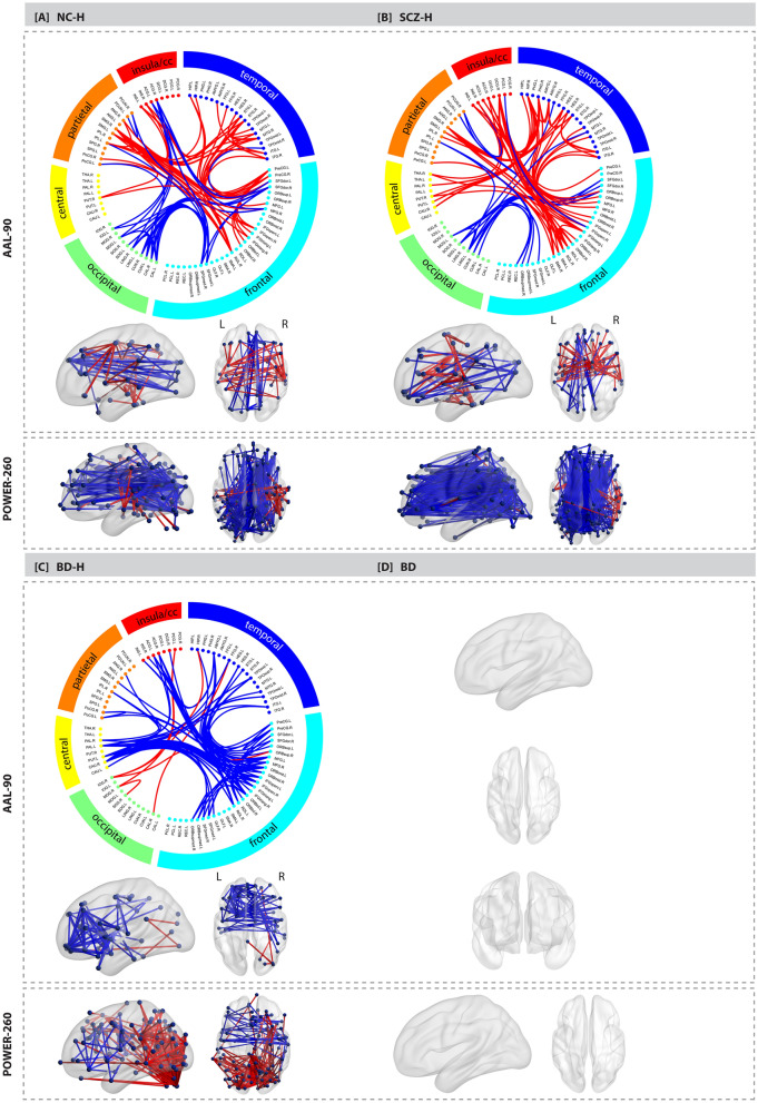
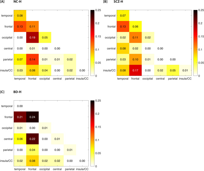
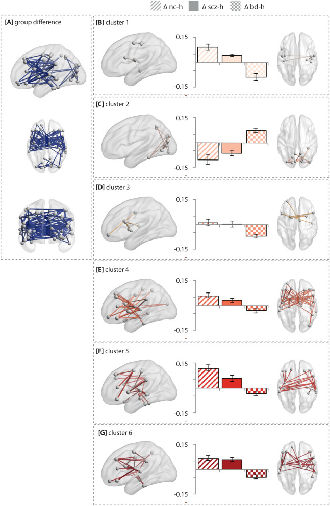
Hallucinations may arise from an imbalance between sensory and higher cognitive brain regions, reflected by alterations in functional connectivity. It is unknown whether hallucinations across the psychosis continuum exhibit similar alterations in functional connectivity, suggesting a common neural mechanism, or whether different mechanisms link to hallucinations across phenotypes. We acquired resting-state functional MRI scans of 483 participants, including 40 non-clinical individuals with hallucinations, 99 schizophrenia patients with hallucinations, 74 bipolar-I disorder patients with hallucinations, 42 bipolar-I disorder patients without hallucinations, and 228 healthy controls. The weighted connectivity matrices were compared using network-based statistics. Non-clinical individuals with hallucinations and schizophrenia patients with hallucinations exhibited increased connectivity, mainly among fronto-temporal and fronto-insula/cingulate areas compared to controls (P < 0.001 adjusted). Differential effects were observed for bipolar-I disorder patients with hallucinations versus controls, mainly characterized by decreased connectivity between fronto-temporal and fronto-striatal areas (P = 0.012 adjusted). No connectivity alterations were found between bipolar-I disorder patients without hallucinations and controls. Our results support the notion that hallucinations in non-clinical individuals and schizophrenia patients are related to altered interactions between sensory and higher-order cognitive brain regions. However, a different dysconnectivity pattern was observed for bipolar-I disorder patients with hallucinations, which implies a different neural mechanism across the psychosis continuum.


Conflict of interest statement

The authors declare no competing interests.

Figures

Figure 1
Figure 1
Functional connectome alterations in the non-clinical and clinical groups compared to healthy controls. The Network Based Statistics results of the AAL atlas are depicted in de upper grey panel and the results of the Power atlas in the lower grey panel: (a) non-clinical individuals with hallucinations (n = 40) vs healthy controls (n = 228); (b) schizophrenia patients with hallucinations (n = 99) vs healthy controls (n = 228); (c) bipolar-I disorder patients with lifetime history of hallucinations (n = 74) vs healthy controls (n = 228). (d) bipolar-I disorder patients without a lifetime history of psychosis (n = 42) did not differ from controls (n = 228). The edges are color-coded based on either an increase (red) or decrease (blue) in connectivity compared to controls. The circle plot depicts all 90 nodes of the AAL atlas clustered according to their cerebral lobe. Group differences were tested at 10,000 permutations P < 0.05 FWE corrected. Age and sex were included as covariates. The corresponding test-statistics, a list of altered connections (Supplementary Tables 6–8 for AAL atlas, Supplementary Tables 10–12 for Power atlas), and abbreviations for the AAL brain regions depicted in the circle plot (Supplementary Table 16) can be found in the Supplementary. BD bipolar-I disorder without lifetime history of hallucinations, BD-H bipolar-I disorder with lifetime history of hallucinations, CC cingulate cortex, NC-H non-clinical individuals with hallucinations, SCZ-H schizophrenia spectrum disorder with hallucinations.
Figure 2
Figure 2
Proportion of altered connections between each pair of lobes for the three hallucination groups relative to controls. The number of altered links between each pair of lobes was divided by the total number of altered pair-wise links based on the results of the AAL atlas. (a) non-clinical individuals with hallucinations (n = 40) vs healthy controls (n = 228); (b) schizophrenia patients with hallucinations (n = 99) vs healthy controls (n = 228); (c) bipolar-I disorder patients with lifetime history of hallucinations (n = 74) vs healthy controls (n = 228). Note that the hippocampus and amygdala are assigned to the temporal lobe. The putamen, pallidum and caudate are included in the central lobe. BD bipolar-I disorder without lifetime history of hallucinations, BD-H bipolar-I disorder with lifetime history of hallucinations, CC cingulate cortex, NC-H non-clinical individuals with hallucinations, SCZ-H schizophrenia spectrum disorder with hallucinations.
Figure 3
Figure 3
Sets of connections clustered according to their behavior in clinical and non-clinical individuals with hallucinations. (A) The result of the overall group comparison between patients with schizophrenia (n = 99), patients with bipolar-I disorder with (n = 74) and non-clinical individuals with hallucinations (n = 40) using the AAL atlas. (B–E) the edges in the component that differed between the groups were clustered by a k-means clustering algorithm to elucidate differences across the groups with hallucinations. Error bars represent the standard deviation of connectivity strength within that particular cluster. The group differences were tested using an F-test at T = 8.0; P < 0.05 at 10,000 permutations. The test-statistics and AAL labels corresponding to the altered connections per cluster can be found in Supplementary Table 9. Age and sex were included as covariates. NC-H non-clinical individuals with hallucinations, BD-H bipolar-I disorder with lifetime history of hallucinations, SCZ-H schizophrenia spectrum disorder with hallucinations.

Similar articles

Cited by

References

    1. Sommer IE, Kahn RS. Psychosis susceptibility syndrome: An alternative name for schizophrenia. Lancet Psychiatry. 2014;1:111. doi: 10.1016/S2215-0366(14)70288-3. - DOI - PubMed
    1. Sommer IE, Koops S, Blom JD. Comparison of auditory hallucinations across different disorders and syndromes. Neuropsychiatry. 2012;2:1–12. doi: 10.2217/npy.12.2. - DOI
    1. Van Os J, Hanssen M, Bijl RV, Ravelli A. Strauss (1969) revisited: A psychosis continuum in the general population? Schizophr. Res. 2000;45:11–20. doi: 10.1016/S0920-9964(99)00224-8. - DOI - PubMed
    1. Jardri R, Pouchet A, Pins D, Thomas P. Cortical activations during auditory verbal hallucinations in schizophrenia: A coordinate-based meta-analysis. Am. J. Psychiatry. 2011;168:73–81. doi: 10.1176/appi.ajp.2010.09101522. - DOI - PubMed
    1. Kompus K, Westerhausen R, Hugdahl K. The, “paradoxical” engagement of the primary auditory cortex in patients with auditory verbal hallucinations: A meta-analysis of functional neuroimaging studies. Neuropsychologia. 2011;49:3361–3369. doi: 10.1016/j.neuropsychologia.2011.08.010. - DOI - PubMed

Publication types

MeSH terms