Review
. 2021 Jun;21(6):347-362.
doi: 10.1038/s41577-020-00477-9. Epub 2021 Jan 13.

Regulation of immune responses by the airway epithelial cell landscape

Affiliations
Review

Regulation of immune responses by the airway epithelial cell landscape

Richard J Hewitt et al. Nat Rev Immunol. 2021 Jun.

Abstract

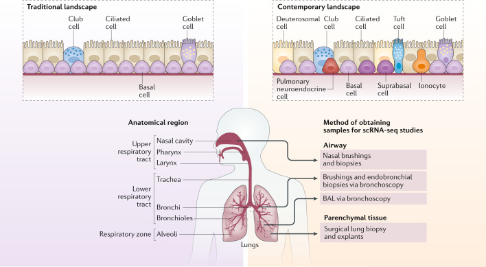
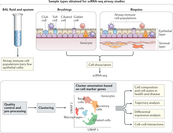
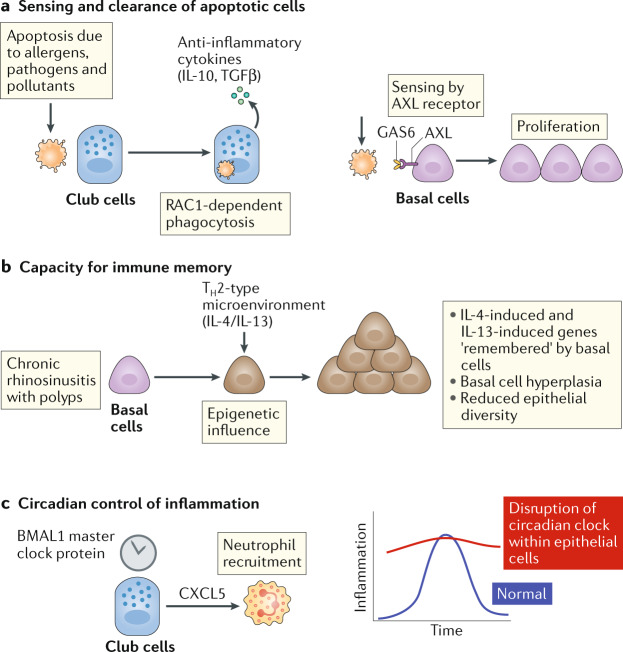
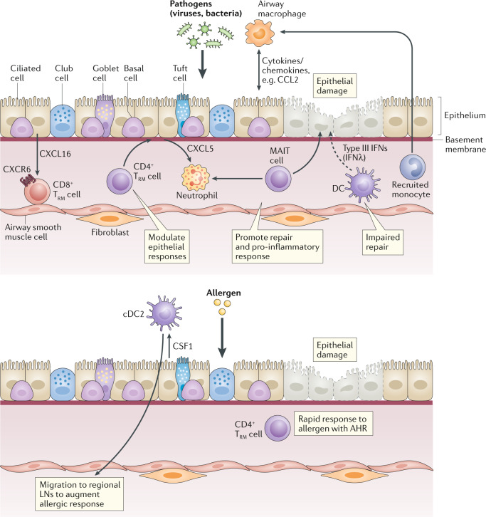
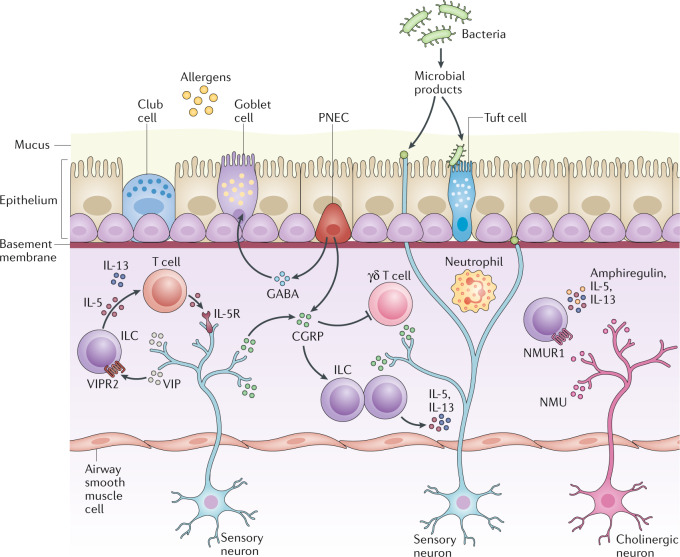
The community of cells lining our airways plays a collaborative role in the preservation of immune homeostasis in the lung and provides protection from the pathogens and pollutants in the air we breathe. In addition to its structural attributes that provide effective mucociliary clearance of the lower airspace, the airway epithelium is an immunologically active barrier surface that senses changes in the airway environment and interacts with resident and recruited immune cells. Single-cell RNA-sequencing is illuminating the cellular heterogeneity that exists in the airway wall and has identified novel cell populations with unique molecular signatures, trajectories of differentiation and diverse functions in health and disease. In this Review, we discuss how our view of the airway epithelial landscape has evolved with the advent of transcriptomic approaches to cellular phenotyping, with a focus on epithelial interactions with the local neuronal and immune systems.


Conflict of interest statement

The authors declare no competing interests.

Figures

Fig. 1
Fig. 1. Revised cell repertoire of human airway epithelium captured by scRNA-seq.
The traditional view of the airway epithelium — comprising epithelial cells lining the upper and lower respiratory tract — has been transformed by single-cell RNA sequencing (scRNA-seq). The revised contemporary landscape features newly identified cell types, such as the ionocyte, and different cell states. The cellular composition and cell states within the airway varies according to anatomical location (proximal–distal axis) and the presence of disease. The approach used to obtain human samples for sequencing differs according to lung compartment; the principle methods for sampling human airway epithelial cells are bronchial brushings and endobronchial biopsies conducted during bronchoscopy, in contrast to sampling of the alveolar region, which is achieved using parenchymal lung tissue obtained from surgical biopsy or from explants. BAL, bronchoalveolar lavage.
Fig. 2
Fig. 2. Single-cell transcriptomic studies of the airway.
For human single-cell RNA sequencing (scRNA-seq) studies of the lower respiratory tract, a flexible bronchoscopy is performed to obtain bronchoalveolar lavage (BAL) fluid, airway wall brushings and endobronchial biopsies. The starting material determines the relative contributions of airway immune cells, epithelial cells and underlying stromal cells that are sequenced. A viable, single-cell suspension is obtained by enzymatic dissociation of brushings and biopsies. Bioinformatic pipelines generate distinct cell clusters by dimensional reduction and visualization techniques such as uniform manifold approximation and projection (UMAP) and facilitate assignment of cluster annotations based on cell-marker genes. Novel cell clusters and cell states can be determined in health and disease and trajectory analysis can be used to study dynamic, differentiating cell types.
Fig. 3
Fig. 3. Immune properties of epithelial cells.
In addition to orchestrating local immune cells through the release of soluble mediators and cell–cell contact, epithelial cells possess intrinsic immune capabilities that directly influence barrier immunity. These include sensing of apoptotic cells by AXL receptor and clearance of apoptotic cells by RAC1 (ref.) (part a), inflammatory memory (part b) and circadian control of inflammation, (part c). BMAL1, brain and muscle ARNT-like 1; CXCL5, CXC-chemokine ligand 5; GAS6, growth arrest-specific protein 6; TH2, T helper 2; TGFβ, transforming growth factor-β.
Fig. 4
Fig. 4. Resident epithelial–immune interactions in the airway.
Complex crosstalk exists between epithelial cells and resident and recruited immune cell populations in the airways. These interactions depend on the specific environmental stimuli, for example, infection with viruses and bacteria or exposure to allergens. Transcriptomic studies have provided enhanced insight into the changes in cell functional state that are driven by these interactions. This figure provides specific examples and is not exhaustive. AHR, airway hyperresponsiveness; cDC2, type 2 conventional dendritic cell; CSF1, colony-stimulating factor 1; CXCL, CXC-chemokine ligand; CXCR6, CXC-chemokine receptor 6; DC, dendritic cell; IFN, interferon; LN, lymph node; MAIT cell, mucosal associated invariant T cell; TRM cell, tissue resident memory T cell.
Fig. 5
Fig. 5. Neuroimmune interactions at the pulmonary epithelium.
The lungs are densely innervated and complex interactions between epithelial cells, neurons and immune cells facilitate sensing of the external inhaled environment. Pulmonary reactions to stimuli, such as pathogens, pollutants, toxins and environmental changes, result in avoidance reflexes and inflammation. Sensory neurons release vasoactive peptide (VIP), which binds to VIP receptor 2 (VIPR2) expressed by group 2 innate lymphoid cells (ILCs) and T helper 2 cells, resulting in the release of IL-5, which stimulates the release of more IL-5 from these neurons, generating an amplifying loop to enhance allergic responses. Communication between group 2 ILCs and pulmonary neuroendocrine cells (PNECs) occurs via calcitonin gene-related peptide (CGRP) to maximize the ILC-derived expression of IL-5 and GABA to elicit mucus generation. By contrast, CGRP inhibits neutrophils and γδ T cells during bacterial infections. Cholinergic neurons react to allergens and helminths to amplify type 2 inflammation via the secretion of neuromedin U (NMU) which reacts with NMU receptor 1 (NMUR1) on group 2 ILCs to enhance cytokine secretion.

Similar articles

Cited by

References

    1. Iwasaki A, Foxman EF, Molony RD. Early local immune defences in the respiratory tract. Nat. Rev. Immunol. 2017;17:7–20. doi: 10.1038/nri.2016.117. - DOI - PMC - PubMed
    1. Rhodin JA. The ciliated cell. Ultrastructure and function of the human tracheal mucosa. Am. Rev. Respir. Dis. 1966;93 (Suppl.):1–15. - PubMed
    1. Breeze RG, Wheeldon EB. The cells of the pulmonary airways. Am. Rev. Respir. Dis. 1977;116:705–777. doi: 10.1164/arrd.1977.116.4.705. - DOI - PubMed
    1. Widdicombe JH. Early studies on the surface epithelium of mammalian airways. Am. J. Physiol. Lung Cell Mol. Physiol. 2019;317:L486–L495. doi: 10.1152/ajplung.00240.2019. - DOI - PubMed
    1. Mercer RR, Russell ML, Roggli VL, Crapo JD. Cell number and distribution in human and rat airways. Am. J. Respir. Cell Mol. Biol. 1994;10:613–624. doi: 10.1165/ajrcmb.10.6.8003339. - DOI - PubMed

Publication types

MeSH terms

Substances