. 2021 Jan 13;11(1):1157.
doi: 10.1038/s41598-021-80961-x.

An in vitro study on Staphylococcus schweitzeri virulence

Affiliations

An in vitro study on Staphylococcus schweitzeri virulence

Almut Grossmann et al. Sci Rep. .

Abstract

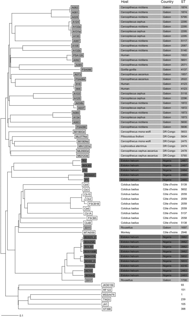
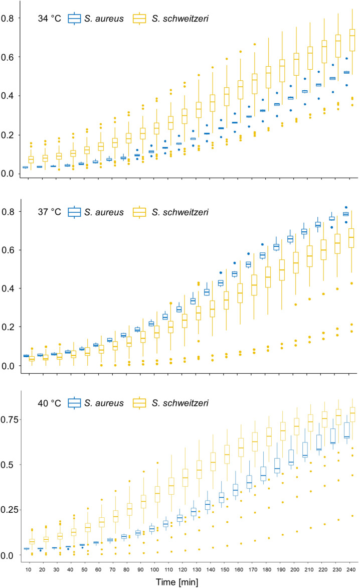
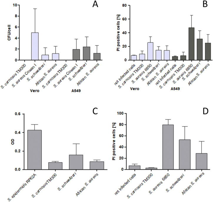
Staphylococcus schweitzeri belongs to the Staphylococcus aureus-related complex and is mainly found in African wildlife; no infections in humans are reported yet. Hence, its medical importance is controversial. The aim of this work was to assess the virulence of S. schweitzeri in vitro. The capacity of African S. schweitzeri (n = 58) for invasion, intra- and extracellular cytotoxicity, phagolysosomal escape, coagulase activity, biofilm formation and host cell activation was compared with S. aureus representing the most common clonal complexes in Africa (CC15, CC121, CC152). Whole genome sequencing revealed that the S. schweitzeri isolates belonged to five geographical clusters. Isolates from humans were found in two different clades. S. schweitzeri and S. aureus showed a similar host cell invasion (0.9 vs. 1.2 CFU/Vero cell), host cell activation (i.e. expression of pro-inflammatory cytokines, 4.1 vs. 1.7 normalized fold change in gene expression of CCL5; 7.3 vs. 9.9 normalized fold change in gene expression of IL8, A549 cells) and intracellular cytotoxicity (31.5% vs. 25% cell death, A549 cells). The extracellular cytotoxicity (52.9% vs. 28.8% cell death, A549 cells) was higher for S. schweitzeri than for S. aureus. Nearly all tested S. schweitzeri (n = 18/20) were able to escape from phagolysosomes. In conclusion, some S. schweitzeri isolates display virulence phenotypes comparable to African S. aureus. S. schweitzeri might become an emerging zoonotic pathogen within the genus Staphylococcus.


Conflict of interest statement

The authors declare no competing interests.

Figures

Figure 1
Figure 1
Neighbour-joining tree of Staphylococcus schweitzeri. The genomes of S. schweitzeri (n = 58) from African wildlife and humans in Côte d’Ivoire, Democratic Republic of the Congo, Nigeria and Gabon were sequenced and the phylogenetic tree was constructed based on the up to 1861 targets of the S. aureus cgMLST scheme. MLST ST, host and country of origin are indicated for each isolate. The colour-code represents the country of origin. S. aureus strains JKD6159, RF122, ST398, JH1, MSSA476 and TW20 were selected for comparison.
Figure 2
Figure 2
Growth curve of Staphylococcus schweitzeri and African Staphylococcus aureus at different temperatures. The growth of S. schweitzeri (n = 58, yellow boxplots) and a representative selection of African S. aureus (n = 6, blue boxplots) was photometrically measures after intervals of 10 min for 4 h. Boxplots (incl. outliers) of optical density (OD) are shown for each species and time points. S. schweitzeri grew faster at 34 °C and 40 °C than S. aureus comparator isolates.
Figure 3
Figure 3
Comparison of the in vitro virulence of Staphylococcus schweitzeri and representative African Staphylococcus aureus. The results were merged for S. schweitzeri (n = 58) and African S. aureus (n = 6) and displayed as the mean (± SD) for each of the two groups. (A) Cellular invasion in Vero and A549 cells. The highly invasive strain S. aureus Cowan I served as positive and the low invasive S. carnosus strain TM300 as negative control. (B) Intracellular cytotoxicity after infection of Vero and A549 cells. The highly cytotoxic S. aureus strain 6850 served as positive and the S. carnosus strain TM300 as negative control. (C) Biofilm formation. The biofilm production was measured photometrically and bars indicated mean OD (± SD). (D) Extracellular cytotoxicity. A549 cells were exposed to secreted toxins in supernatants of overnight cultures. The mean cytotoxicity (proportion of dead cells, ± SD) of S. schweitzeri was significantly stronger than for African S. aureus (p = 0.02).

References

    1. Tong SY, et al. Novel staphylococcal species that form part of a Staphylococcus aureus-related complex: The non-pigmented Staphylococcus argenteus sp. nov. and the non-human primate-associated Staphylococcus schweitzeri sp. nov. Int. J. Syst. Evol. Microbiol. 2015;65:15–22. doi: 10.1099/ijs.0.062752-0. - DOI - PMC - PubMed
    1. Becker K, et al. Implications of identifying the recently defined members of the Staphylococcus aureus complex S. argenteus and S. schweitzeri: A position paper of members of the ESCMID Study Group for Staphylococci and Staphylococcal Diseases (ESGS) Clin. Microbiol. Infect. 2019;1:1. doi: 10.1016/j.cmi.2019.02.028. - DOI - PubMed
    1. Shittu AO, Mellmann A, Schaumburg F. Molecular characterization of Staphylococcus aureus complex from fomites in Nigeria. Infect. Genet. Evol. 2020;85:104504. doi: 10.1016/j.meegid.2020.104504. - DOI - PubMed
    1. Schaumburg F, et al. Staphylococcus aureus complex from animals and humans in three remote African regions. Clin. Microbiol. Infect. 2015;21(345):e341–348. doi: 10.1016/j.cmi.2014.12.001. - DOI - PubMed
    1. Ngoa UA, et al. Epidemiology and population structure of Staphylococcus aureus in various population groups from a rural and semi urban area in Gabon, Central Africa. Acta Trop. 2012;124:42–47. doi: 10.1016/j.actatropica.2012.06.005. - DOI - PubMed

Publication types

Supplementary concepts

LinkOut - more resources