. 2021 Feb;590(7847):666-670.
doi: 10.1038/s41586-020-03124-4. Epub 2021 Jan 13.

Functional refolding of the penetration protein on a non-enveloped virus

Affiliations

Functional refolding of the penetration protein on a non-enveloped virus

Tobias Herrmann et al. Nature. 2021 Feb.

Abstract

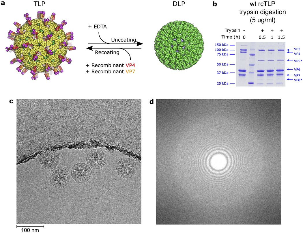
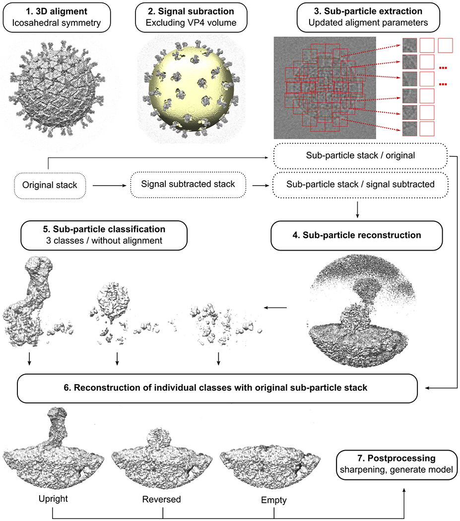
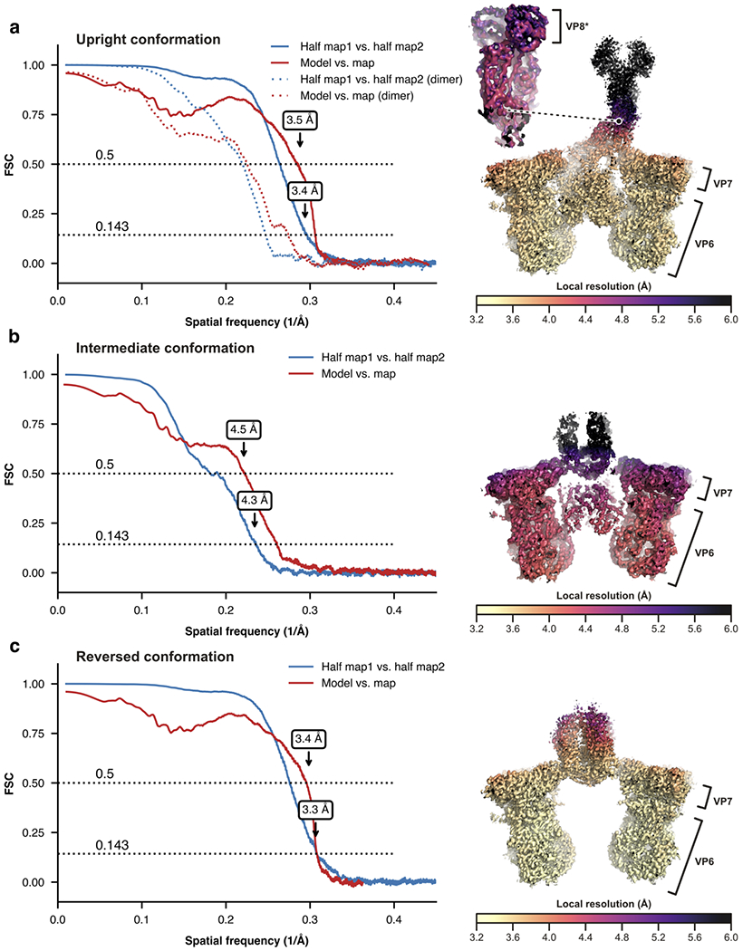
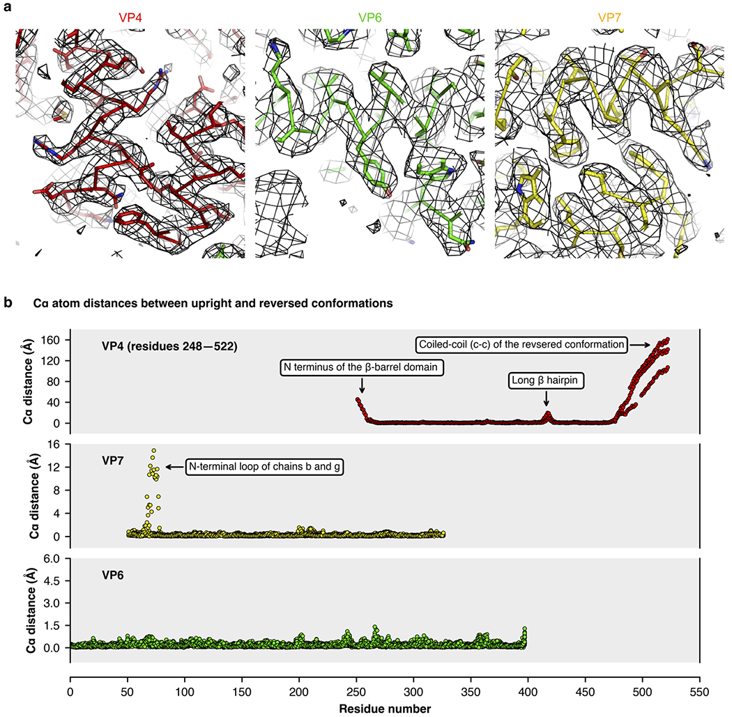
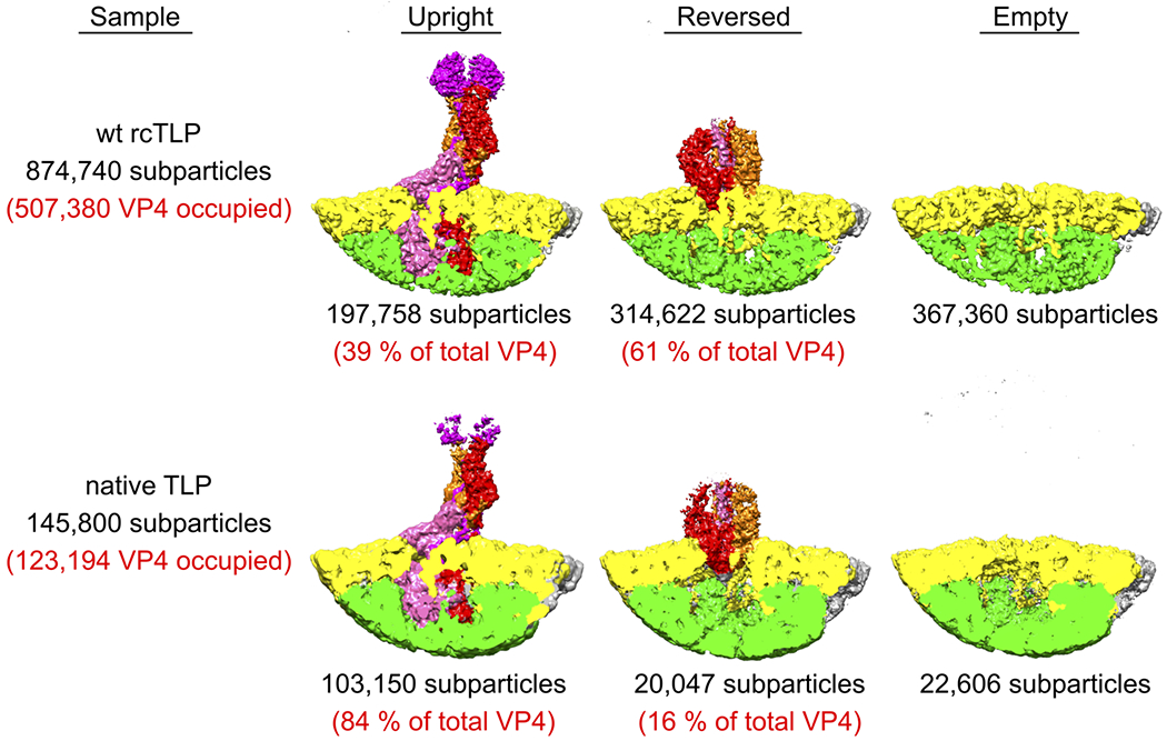
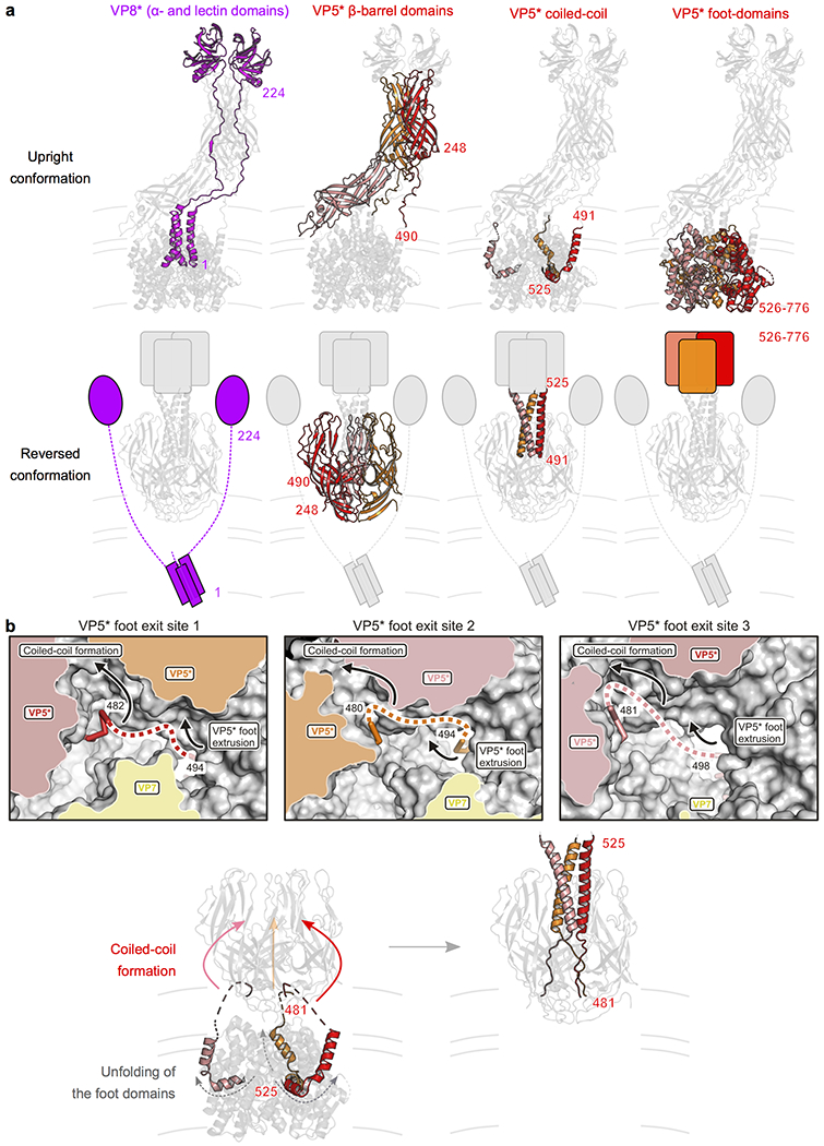
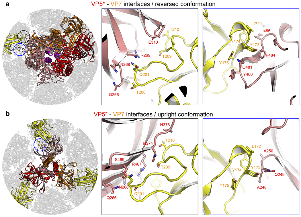
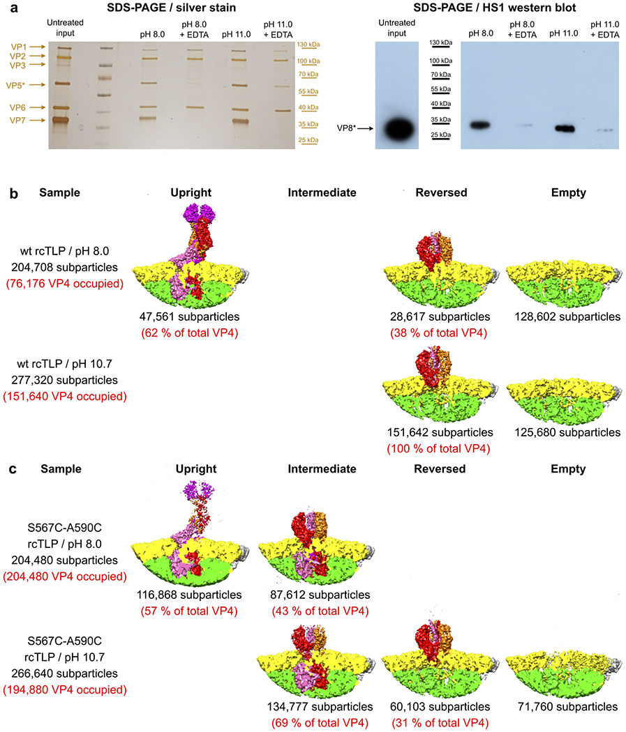
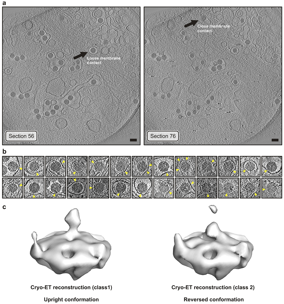
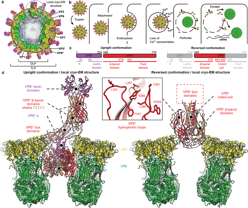
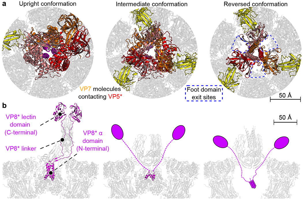
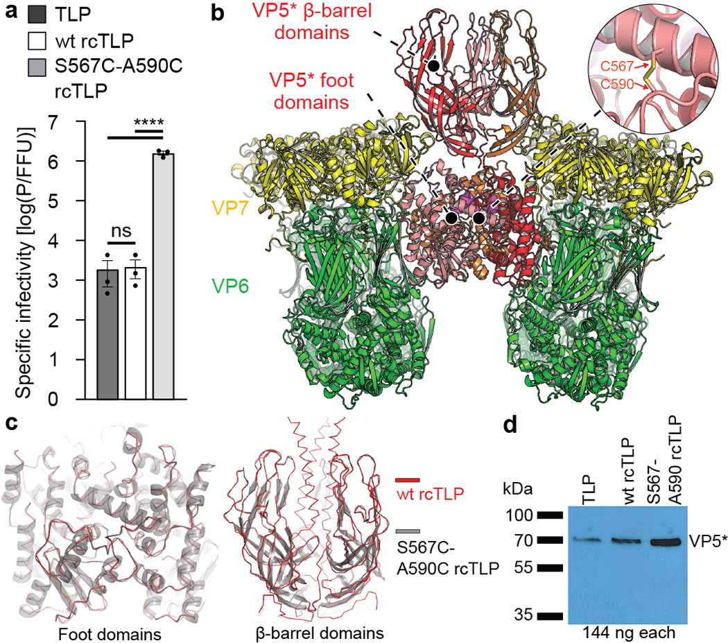
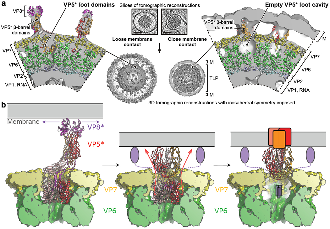
A non-enveloped virus requires a membrane lesion to deliver its genome into a target cell1. For rotaviruses, membrane perforation is a principal function of the viral outer-layer protein, VP42,3. Here we describe the use of electron cryomicroscopy to determine how VP4 performs this function and show that when activated by cleavage to VP8* and VP5*, VP4 can rearrange on the virion surface from an 'upright' to a 'reversed' conformation. The reversed structure projects a previously buried 'foot' domain outwards into the membrane of the host cell to which the virion has attached. Electron cryotomograms of virus particles entering cells are consistent with this picture. Using a disulfide mutant of VP4, we have also stabilized a probable intermediate in the transition between the two conformations. Our results define molecular mechanisms for the first steps of the penetration of rotaviruses into the membranes of target cells and suggest similarities with mechanisms postulated for other viruses.


Conflict of interest statement

Competing interests

The authors declare no competing interests.

Figures

Extended Data Fig. 1 |
Extended Data Fig. 1 |. Sample preparation and cryo-EM data collection.
a, Schematic protocol for recoating of double-layer particles (DLPs) with recombinant VP4 and VP7. b, Time course of wild-type recoated triple-layer particles (wt rcTLPs) trypsin digestion with 5 μg/ml trypsin at 37 °C. Samples were analyzed after the shown incubation times by sodium dodecyl sulfate-polyacrylamide gel electrophoresis (SDS-PAGE). The experiment was repeated independently two times with similar results. For gel source data, see Supplementary Fig. 1. c, Representative micrograph (aligned and summed movie frames) of wt rcTLPs recorded with a Polara F30 electron microscope equipped with a K2 summit detector (magnification = 40650). The scale bar corresponds to 100 nm. d, Power spectrum of the micrograph shown in c.
Extended Data Fig. 2 |
Data processing workflow for local reconstructions of rotavirus spike proteins.
Extended Data Fig. 3 |
Extended Data Fig. 3 |. Resolution analysis of the local cryo-EM reconstructions.
Left, Fourier shell correlation (FSC) curves for the reconstructions and refined models calculated with phenix.mtriage. Correlations for the two half maps are shown as solid blue lines after applying a mask encompassing the models. Correlations between the refined model and final map are shown as red solid lines. Nominal resolution estimates at conventional FSC values are indicated by arrows. Right, local resolution of the reconstructions calculated with ResMap. a, Upright conformation. Dashed lines (left) are the FSC analysis for the reconstruction of the distal VP5*/VP8* dimeric density that was obtained through alignment by classification (see Methods). The inset (right) shows the local resolution of this reconstruction. b, Intermediate conformation. c, Reversed conformation. For source data of the FCS plots, see Supplementary Data 5.
Extended Data Fig. 4 |
Extended Data Fig. 4 |. cryo-EM density and structure comparison.
a, Close-up views of representative regions of the cryo-EM density maps obtained by local reconstruction. Density is shown as gray mesh; polypeptide-chain backbone as ribbon; side-chain atoms as sticks (carbon, black; nitrogen, blue; oxygen, red; sulfur, orange). b, Per-residue Cα distances after subunit-vise superposition of V4, VP7, and VP6 subunits from the upright and reversed conformation structures.
Extended Data Fig. 5 |
Extended Data Fig. 5 |. Comparison of penetration protein conformations on rcTLPs and native TLPs.
Relative subparticle amounts, and corresponding cryo-EM reconstructions obtained from rcTLPs (top row) and native TLPs (bottom row). Local spike reconstructions of rcTLPs were obtained from two cryo-EM samples prepared from two independent recoating reactions. Local spike reconstructions of native TLPs were obtained from one cryo-EM sample.
Extended Data Fig. 6 |
Extended Data Fig. 6 |. Rearrangements of the VP8* and VP5* penetration proteins during transition from upright to reversed conformation on the virion surface.
a, Distinct domains of the VP8* and VP5* spike proteins are colored separately to illustrate their conformational change during transition from upright (top row) to reversed (bottom row) conformation. Domains that were not observed in our cryo-EM maps because of flexible attachment are drawn schematically. VP8*, magenta; VP5*, red, orange and salmon. b, Formation of the trimeric coiled coil and extrusion of the foot domains. Top row, close-up views of the VP5* foot domain exit sites as observed in the intermediate conformation structure. A partially cut surface representation is shown. VP5*, red, orange and salmon; VP7, yellow. The last modeled residues of the VP5* β-barrel domains, 482 (chain 1), 480 (chain 2), and 481 (chain 3), are located on the outside. The connections to the first modeled VP5* foot domain residue, 494 (chain 1), 494 (chain 2), and 498 (chain 3), are indicated by dashed lines (fuzzy density in the cryo-EM map). Arrows indicate a suggested mechanism for foot domain reversal, involving zipping-up of the trimeric coiled-coil and unfolding and extrusion of foot domain residues. Bottom row, proposed transition from the intermediate structure (left) to the reversed structure by “zipping up” of the coiled coil and unfolding and extrusion of the foot domains.
Extended Data Fig. 7 |
Extended Data Fig. 7 |. Molecular details of the VP5*-VP7 interfaces for the upright and reversed conformations.
VP5*, salmon; VP7, yellow. a, VP5*-VP7 interface for the VP5* upright conformation. b, VP5*-VP7 interface for the VP5* reversed conformation. The following VP5* interface residues are conserved (Supplementary Data 1): N268, N376, R467, S469. The VP5* β-barrel N terminus (residues 248–250) is not strictly conserved, however, the interaction is based on main-chain hydrogen bonds and can likely be maintained for different side chains as well. The following VP7 interface residues are conserved (Supplementary Data 2): S201, T210, L172–Y175.
Extended Data Fig. 8 |
Extended Data Fig. 8 |. Inducing penetration protein reversal at alkaline pH and VP8* association with TLPs.
a, Analysis of rotavirus particles without (lanes 1 and 2) and with (lines 3 and 4) pH-induced conformational change of VP8*/VP5*, and without (lanes 1 and 3) and with (lanes 2 and 4) EDTA-induced uncoating. Pelleted fractions were analyzed by SDS-PAGE and silver staining (left) and by Western blotting with the VP8*-specific antibody HS1 (right) (see Methods). The experiment was repeated independently three times with similar results. For gel and Western blot source data, see Supplementary Fig. 1. b, Relative VP4 subparticle amounts, and corresponding cryo-EM reconstructions obtained from wild-type (wt) rcTLPs. c, Relative VP4 subparticle amounts, and corresponding cryo-EM reconstructions obtained from S567C-A590C rcTLPs. Recoating reactions for all samples were carried out at the same time and with the same VP7 and DLP stock solutions. All cryo-EM samples were prepared in the same blotting session.
Extended Data Fig. 9 |
Extended Data Fig. 9 |. Cryo-tomographic (cryo-ET) analysis of RRVs entering BSC-1 cells.
a, Sections of the tomogram from which the reconstructions (icosahedral average of single virion subtomograms) in Fig. 4a were obtained. Left, virus with “loose” membrane contact indicated by an arrow. Right, virus with “tight” membrane contact indicated by an arrow. Images were low-pass filtered and contrast enhanced for display. Scale bar corresponds to 100 nm. b, Tomographic slices of manually selected viruses (not including particle in panel a) from several tomograms with tight membrane contacts (yellow arrowheads). Images were low-pass filtered and contrast enhanced for display. Those particles were selected to detect VP4 reversed conformations in additional viruses (other than particle in Fig. 4a). c, Sub-subtomogram classification of VP4 positions extracted from the RRV particles shown in panel b yielded a class (left) with the upright conformation and a class (right) with the reversed conformation. VP4 positions were extracted from 26 selected particles chosen to have an easily identified region of close membrane contact. The particle shown in Fig. 4a was excluded from this selection. Note that the reconstructions shown in Fig. 4a are from single particles with imposed icosahedral symmetry, whereas we show here classified averaged individual volumes extracted at VP4 positions.
Fig. 1 |
Fig. 1 |. Rotavirus entry and cryo-EM structures of the penetration protein in upright and reversed conformations.
a, Overall structure of a rotavirus TLP. b, Schematic virus entry pathway derived from live-cell imaging experiments,,. c, Domain organization of the VP8* and VP5* proteins in the upright (left) and reversed (right) conformations. Domains are labeled beneath the primary structure. Residue numbers indicate termini and domain boundaries; α, N-terminal α-helical segment of VP8*; regions in grey, not detected (disordered) in the reversed-conformation structure. d, Atomic models of upright (left) and reversed (right) conformations of VP5* (and VP8*) on the virion surface, from our cryo-EM reconstructions of wild-type recoated RRV TLPs. VP5*, red, orange and salmon; VP6, green; VP7, yellow; VP8*, magenta. Some VP6 and VP7 subunits omitted for clarity.
Fig. 2 |
Fig. 2 |. Interactions of the penetration protein domains VP5* and VP8* with VP7 capsid subunits.
a, Top views of the upright (left), intermediate (middle), and reversed (right) conformations. VP5*, red, orange and salmon; VP7 chains interacting with VP5*, yellow. Close-up views of the interfaces are in Extended Data Fig. 7. Gaps between VP5* and VP7 shell molecules that allow for exit of VP5* foot domains are marked by blue dashed boxes. b, Side views of the upright (left), intermediate (middle) and reversed (right) conformations. VP8*, magenta. In the reversed structure (right), VP8* is shown schematically: no density for it was observed, but biochemical data shows its presence.
Fig. 3 |
Fig. 3 |. Infectivity and structure of TLPs recoated with mutant S567C-A590C penetration proteins.
a, Particle to focus-forming unit (FFU) ratios of TLPs, wt rcTLPs and S567C-A590C rcTLPs determined as described in Methods. n = 3 biologically independent experiments. Error bars, mean ± s.d. Statistics: one-way ANOVA with Tukey post hoc test. p < 0.0001 for overall test (**** = p < 0.0001, ns = not significant). For source data, see Supplementary Data 4. b, Structure of the foot-locked intermediate obtained from cryo-EM analysis of S567C-A590C rcTLPs. VP5*, red, orange and salmon; VP6, green; VP7, yellow; VP8*, magenta. c, Left, structural comparison of the VP5* foot domains from the upright conformation of wt rcTLPs (red) and intermediate conformation of S567C-A590C rcTLPs (grey). Right, structural comparison of VP5* β-barrel domains from the reversed conformation of wt rcTLPs (red) and intermediate conformation of S567C-A590C rcTLPs (grey). d, Comparison of relative amounts of VP5* determined by Western blotting as described in Methods. Experiment repeated independently two times with similar results. For Western blot source data, see Supplementary Fig. 1.
Fig. 4 |
Fig. 4 |. Molecular rearrangements of the membrane-penetration protein during rotavirus entry.
a, Cryo-ET reconstructions of RRV entering at the thin edge of BSC-1 cells. Tomographic analysis is shown for two viral particles that are representative for a “loose” (left) or “tight” (right) virion-membrane (M) interface, respectively. In the close-up views of the tomographic reconstructions, density is partially cut and shown in solid gray; protein subunits of positioned atomic models are shown in ribbon representation and colored as in Fig. 1. The scale bar in the tomographic slices (low-pass filtered and contrast-enhanced) is 50 nm. b, Model for VP5* and VP8* rearrangement and membrane-interaction; see Discussion for details.

Similar articles

Cited by

References

    1. Harrison SC in Fields Virology (6th edition) (ed Knipe DM and Howley PM) 52–86 (Wolters Kluwer/Lippincott Williams and Wilkins, 2013).
    1. Estes MK & Greenberg H in Fields Virology (6th edition) (ed Knipe DM and Howley PM) 1347–1401 (Wolters Kluwer/Lippincott Williams and Wilkins, 2013).
    1. Trask SD, Ogden KM & Patton JT Interactions among capsid proteins orchestrate rotavirus particle functions. Curr. Opin. Virol 2, 373–379, doi:10.1016/j.coviro.2012.04.005 (2012). - DOI - PMC - PubMed
    1. Tihova M, Dryden KA, Bellamy AR, Greenberg HB & Yeager M Localization of membrane permeabilization and receptor binding sites on the VP4 hemagglutinin of rotavirus: implications for cell entry. J. Mol. Biol 314, 985–992, doi:10.1006/jmbi.2000.5238 (2001). - DOI - PubMed
    1. Kim IS, Trask SD, Babyonyshev M, Dormitzer PR & Harrison SC Effect of mutations in VP5 hydrophobic loops on rotavirus cell entry. J. Virol 84, 6200–6207, doi:10.1128/JVI.02461-09 (2010). - DOI - PMC - PubMed

Publication types

MeSH terms

LinkOut - more resources