Case Reports
. 2018;33(1):49-52.
doi: 10.15605/jafes.033.01.08. Epub 2018 Apr 20.

Communicating Hydrocephalus in a Case of Long-Term Primary Hyperparathyroidism

Affiliations
Case Reports

Communicating Hydrocephalus in a Case of Long-Term Primary Hyperparathyroidism

Cheow Peng Ooi et al. J ASEAN Fed Endocr Soc. 2018.

Abstract

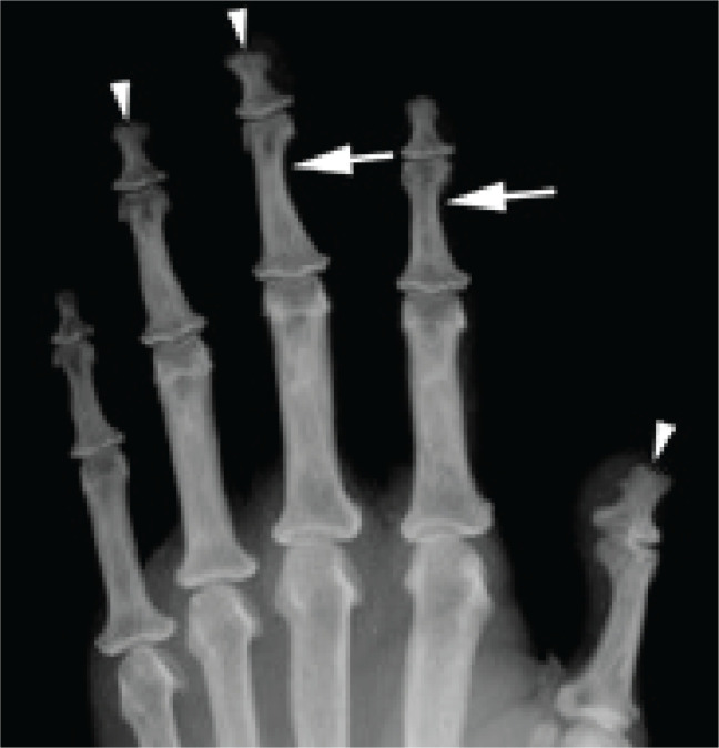
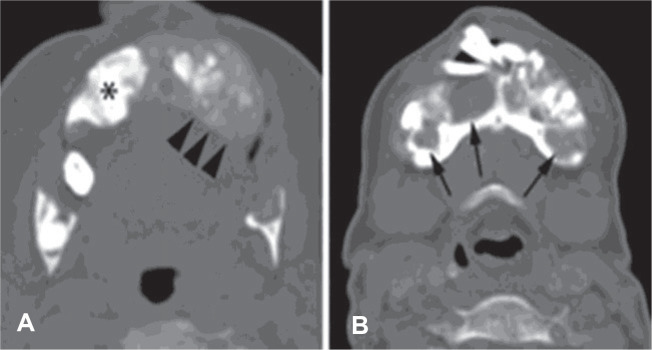
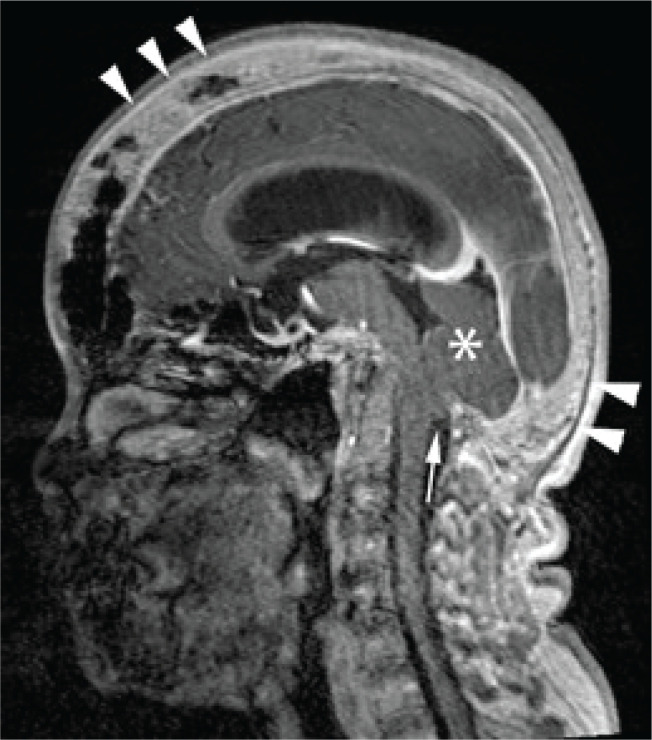
We present the rare case of a 47-year-old woman with protracted primary hyperparathyroidism complicated by communicating hydrocephalus and cerebellar tonsillar herniation secondary to calvarial thickening. The parathyroid glands remained elusive, despite the use of advanced preoperative imaging modalities and three neck explorations. The serum calcium was optimally controlled with cinacalcet and alfacalcidol. Awareness of this rare complication is essential for early diagnosis and prompt intervention to prevent fatal posterior brain herniation.

Keywords: cinacalcet; hydrocephalus; hypercalcemia; hyperparathyroidism; osteitis fibrosis cystica; primary.


Conflict of interest statement

The authors declared no conflict of interest.

Figures

Figure 1
Figure 1
AP radiograph of the left hand, cropped to accentuate osseous detail. Areas of subperiosteal resorption are seen markedly at the radial aspect of the third middle phalanx, and subtly at the radial aspect of the second middle phalanx (arrows). Acro-osteolysis is also evident at the distal phalanges (arrowheads).
Figure 2
Figure 2
Cross-sectional CT of the mandible in bone algorithm at the level of the mandibular angles (A) and symphysis menti (B). There is a loss of cortical and marrow differentiation. This has been replaced by a ground glass density, with areas of irregular osseous expansion (arrowheads in A) and patchy regions of osteosclerosis (asterisk in A). Cystic lesions with wellcircumscribed sclerotic margins (arrows in B) are likely Brown tumours, with evident dental displacement.
Figure 3
Figure 3
T1 post-gadolinium MRI in midsagittal reconstruction. Calvarial thickening, a known effect of hyperparathyroidism, is apparent (arrowheads). Note the ventricular dilatation and the small posterior fossa (asterisk). Cerebellar tonsillar herniation is depicted (arrow).

Similar articles

References

    1. Singh Ospina NM, Rodriguez-Gutierrez R, Maraka S et al. . Outcomes of parathyroidectomy in patients with primary hyperparathyroidism: A systematic review and meta-analysis. World J Surg. 2016;40(10): 2359-77. PMID: 10.1007/s00268-016-3514-1 - DOI - PubMed
    1. Guerin C, Paladino NC, Lowery A, Castinetti F, Taieb D, Sebag F. Persistent and recurrent hyperparathyroidism. Updates Surg. 2017;69(2):161-9. PMID: 10.1007/s13304-017-0447-7 - DOI - PubMed
    1. Robinson PJ, Woodhead P. Primary hyperparathyroidism presenting with a maxillary tumour and hydrocephalus. J Laryngol Otol. 1988;102(12):1164-7. PMID: . - PubMed
    1. Brabyn P, Capote A, Belloti M, Zylberberg I. Hyperparathyroidism diagnosed due to brown tumors of the jaw: A case report and literature review. J Oral Maxillofac Surg. 2017;75(10):2162-9. PMID: 10.1016/j.joms.2017.03.013 - DOI - PubMed
    1. Al-Gahtany M, Cusimano M, Singer W, Bilbao J, Kovacs K, Marotta T. Brown tumors of the skull base. Case report and review of the literature. J Neurosurg. 2003;98(2):417-20. PMID: 10.3171/jns.2003.98.2.0417 - DOI - PubMed

Publication types

LinkOut - more resources