Review
. 2021 Jan;37(1):70-77.
doi: 10.1007/s12055-020-00997-y. Epub 2020 Jul 21.

Use of the hybrid room for thoracic surgery procedures: single-stage localization and removal of non-palpable nodules

Affiliations
Review

Use of the hybrid room for thoracic surgery procedures: single-stage localization and removal of non-palpable nodules

Giulio Melloni et al. Indian J Thorac Cardiovasc Surg. 2021 Jan.

Abstract

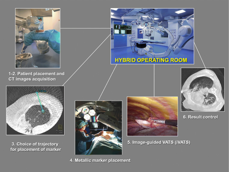
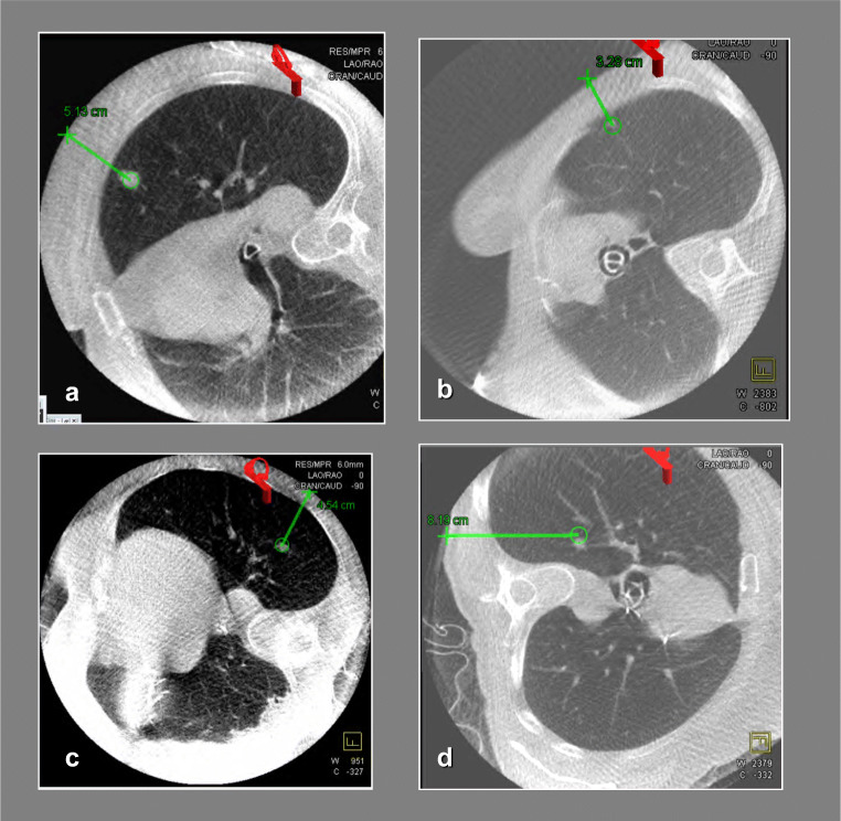
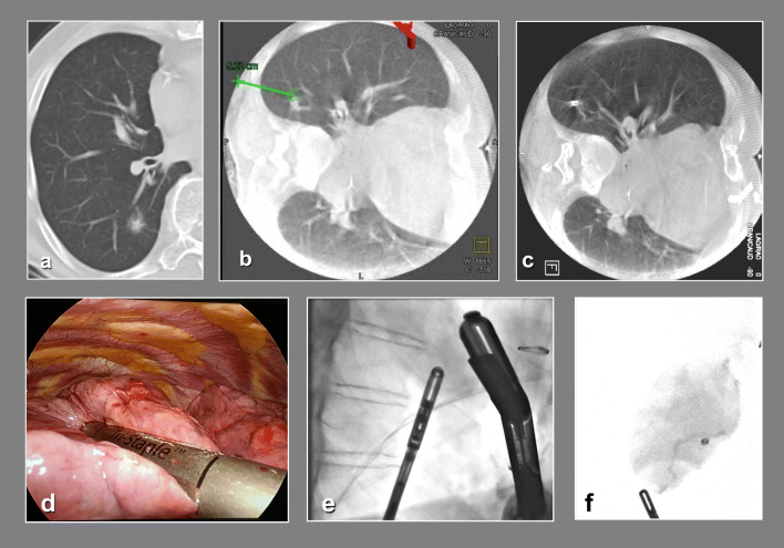
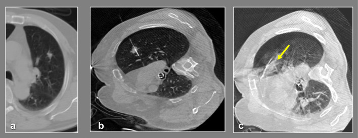
With the widespread availability of lung cancer screening programs, the number of small lung nodules requiring histological characterization has dramatically increased. Because computed tomography-guided fine-needle aspiration may frequently yield false-negative results, excisional biopsy using thoracoscopy is frequently required. Although thoracoscopic procedure has been known to be ideal for nodule resection, the identification of very small, subsolid and deep pulmonary nodules may still be challenging. Precise lesion localization is a key prerequisite to avoid conversion to an unplanned thoracotomy. In the traditional workflow, the localization procedure is performed in the radiology suite, after which the patient is moved to an operating room. With the availability of hybrid operating rooms, a new approach encompassing simultaneous localization and removal of non-palpable lung nodules has become feasible. In this article, we review the procedural workflow of this new technique and discuss its indications and results.

Keywords: Hybrid operating room; Image-guided video–assisted thoracoscopic surgery (iVATS); Localization; Lung nodule.


Conflict of interest statement

Conflict of interestThe authors declare that they have no conflict of interest.

Figures

Fig. 1
Fig. 1
The procedural workflow of single-stage localization and removal of non-palpable lung nodules through iVATS, articulated into 6 steps (see text)
Fig. 2
Fig. 2
Criteria for marker selection. a, b A hookwire is preferred if the marker can be placed with a trajectory perpendicular to the nearest pleural surface (with a deviation of ± 30°). If that is impossible, an anatomical obstacle exists (e.g. scapula) or the lesion is very deep, a coil is used (c, d)
Fig. 3
Fig. 3
a The preoperative chest CT scan showing a subsolid lesion of the apical segment of the right lower lobe. In the b, the needle trajectory is illustrated. The coil was inserted and the marker final position was checked with CBCT (c). During iVATS (d), fluoroscopy allowed intraoperative localization of the coil (e). The resection was performed with endostaplers. Fluoroscopy was also used to confirm that the coil was within the specimen (f)
Fig. 4
Fig. 4
a Preoperative CT scan of the chest. b Intraoperative cone-beam CT scan performed after the hookwire placement. c Intraoperative cone-beam CT scan performed at the end of VATS resection (see text)

References

    1. Gill RR, Zheng Y, Barlow JS, et al. Image-guided video assisted thoracoscopic surgery (iVATS) – phase I-II clinical trial. J Surg Oncol. 2015;112:18–25. doi: 10.1002/jso.23941. - DOI - PMC - PubMed
    1. Zhao ZR, Lau RWH, Yu PSY, Ng CSH. Devising the guidelines. The techniques of pulmonary nodule localization in uniportal video-assisted thoracic surgery-hybrid operating room in the future. J Thorac Dis. 2019;11:S2073–S2078. doi: 10.21037/jtd.2019.01.82. - DOI - PMC - PubMed
    1. Park CH, Han K, Hur J, et al. Comparative effectiveness and safety of preoperative lung localization for pulmonary nodules. A systematic review and meta-analysis. Chest. 2017;151:316–328. doi: 10.1016/j.chest.2016.09.017. - DOI - PubMed
    1. Stanzi A, Mazza F, Lucio F, et al. Tailored intraoperative localization of non-palpable pulmonary lesions for thoracoscopic wedge resection using hybrid room technology. Clin Respir J. 2018;12:1661–1667. doi: 10.1111/crj.12725. - DOI - PubMed
    1. Hsieh MJ, Wen CT, Fang HY, Wen YW, Lin CC, Chao YK. Learning curve of image-guided video-assisted thoracoscopic surgery for small pulmonary nodules: a prospective analysis of 30 initial patients. J Thorac Cardiovasc Surg. 2018;155:1825–32.e1. doi: 10.1016/j.jtcvs.2017.11.079. - DOI - PubMed

LinkOut - more resources