A Literature Review of Dorsal Root Entry Zone Complex (DREZC) Lesions: Integration of Translational Data for an Evolution to More Accurate Nomenclature
- PMID: 33442287
- PMCID: PMC7800708
- DOI: 10.2147/JPR.S255726
A Literature Review of Dorsal Root Entry Zone Complex (DREZC) Lesions: Integration of Translational Data for an Evolution to More Accurate Nomenclature
Abstract
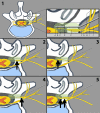
The purpose of this translational review was to provide evidence to support the natural evolution of the nomenclature of neuromodulatory and neuroablative radiofrequency lesions for pain management from lesions of individualized components of the linear dorsal afferent pathway to "Dorsal Root Entry Zone Complex (DREZC) lesions." Literature review was performed to collate anatomic and procedural data and correlate these data to clinical outcomes. There is ample evidence that the individual components of the DREZC (the dorsal rami and its branches, the dorsal root ganglia, the dorsal rootlets, and the dorsal root entry zone) vary dramatically between vertebral levels and individual patients. Procedurally, fluoroscopy, the most commonly utilized technology is a 2-dimensional x-ray-based technology without the ability to accurately locate any one component of the DREZC dorsal afferent pathway, which results in clinical inaccuracies when naming each lesion. Despite the inherent anatomic variability and these procedural limitations, the expected poor clinical outcomes that might follow such nomenclature inaccuracies have not been shown to be prominent, likely because these are all lesions of the same anatomically linear sensory pathway, the DREZC, whereby a lesion in any one part of the pathway would be expected to interrupt sensory transmission of pain to all subsequent more proximal segments. Given that the common clinically available tools (fluoroscopy) are inaccurate to localize each component of the DREZC, it would be inappropriate to continue to erroneously refer to these lesions as lesions of individual components, when the more accurate "DREZC lesions" designation can be utilized. Hence, to avoid inaccuracies in nomenclature and until more accurate imaging technology is commonly utilized, the evidence herein supports the proposed change to this more sensitive and inclusive nomenclature, "DREZC lesions."
Keywords: DREZ; chronic pain; ganglia; pulsed radiofrequency treatment; radiofrequency ablation; spinal.
© 2021 Visnjevac et al.
Conflict of interest statement
Alaa Abd-Elsayed reports serving as a consultant for Medtronic and Avanos. The authors report no other potential conflicts of interest in this body of work.
Figures


Similar articles
-
Safety of Conventional and Pulsed Radiofrequency Lesions of the Dorsal Root Entry Zone Complex (DREZC) for Interventional Pain Management: A Systematic Review.Pain Ther. 2022 Jun;11(2):411-445. doi: 10.1007/s40122-022-00378-w. Epub 2022 Apr 17. Pain Ther. 2022. PMID: 35434768 Free PMC article. Review.
-
Dorsal root entry zone lesions for conus medullaris root avulsions.Appl Neurophysiol. 1988;51(2-5):198-205. doi: 10.1159/000099963. Appl Neurophysiol. 1988. PMID: 3389796
-
Correlation of preoperative MRI with the long-term outcomes of dorsal root entry zone lesioning for brachial plexus avulsion pain.J Neurosurg. 2016 May;124(5):1470-8. doi: 10.3171/2015.2.JNS142572. Epub 2015 Sep 25. J Neurosurg. 2016. PMID: 26406799
-
[Biomechanical characteristics of spinal cord tissue--basis for the development of modifications of the DREZ (dorsal root entry zone) operation].Acta Chir Iugosl. 2004;51(4):59-64. doi: 10.2298/aci0404059s. Acta Chir Iugosl. 2004. PMID: 16018411 Serbian.
-
[Surgery in the dorsal root entry zone for treatment of chronic pain].Neurochirurgie. 2000 Nov;46(5):429-46. Neurochirurgie. 2000. PMID: 11084476 Review. French.
Cited by
-
Safety of Conventional and Pulsed Radiofrequency Lesions of the Dorsal Root Entry Zone Complex (DREZC) for Interventional Pain Management: A Systematic Review.Pain Ther. 2022 Jun;11(2):411-445. doi: 10.1007/s40122-022-00378-w. Epub 2022 Apr 17. Pain Ther. 2022. PMID: 35434768 Free PMC article. Review.
-
Review of Opioid Sparing Interventional Pain Management Options and Techniques for Radiofrequency Ablations for Sacroiliac Joint Pain.Curr Pain Headache Rep. 2022 Nov;26(11):855-862. doi: 10.1007/s11916-022-01088-w. Epub 2022 Sep 30. Curr Pain Headache Rep. 2022. PMID: 36178572 Review.
-
DREZotomy in the era of minimally invasive interventions for cancer-related pain management.Ann Med Surg (Lond). 2024 Jun 17;86(8):4327-4332. doi: 10.1097/MS9.0000000000002288. eCollection 2024 Aug. Ann Med Surg (Lond). 2024. PMID: 39118767 Free PMC article. No abstract available.
-
Radiofrequency Ablation of the Superior Cluneal Nerve: A Novel Minimally Invasive Approach Adopting Recent Anatomic and Neurosurgical Data.Pain Ther. 2022 Jun;11(2):655-665. doi: 10.1007/s40122-022-00385-x. Epub 2022 Apr 17. Pain Ther. 2022. PMID: 35430676 Free PMC article.
References
-
- Pizzo P. Relieving Pain in America: A Blueprint for Transforming Prevention, Care, Education, and Research. Washington (DC); 2011. - PubMed
-
- Purves D, Augustine GJ, Fitzpatrick D. The internal anatomy of the spinal cord In: Purves DAG, Fitzpatrick D, editors. Neuroscience. Sunderland (MA): Sinauer Associates; 2001.
-
- Anatomy and physiology of the spinal cord; 2000–2013. Available from: https://www.ncbi.nlm.nih.gov/books/NBK6229/. Accessed December7, 2020.
-
- Karatas A, Caglar S, Savas A, Elhan A, Erdogan A. Microsurgical anatomy of the dorsal cervical rootlets and dorsal root entry zones. Acta Neurochir (Wien). 2005;147(2):195–199. - PubMed
Publication types
LinkOut - more resources
Full Text Sources
Other Literature Sources

