. 2021 Jan 1;12(4):1200-1211.
doi: 10.7150/jca.54007. eCollection 2021.

LncRNA FAM83A-AS1 promotes ESCC progression by regulating miR-214/CDC25B axis

Affiliations

LncRNA FAM83A-AS1 promotes ESCC progression by regulating miR-214/CDC25B axis

Jinlin Jia et al. J Cancer. .

Abstract

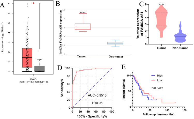
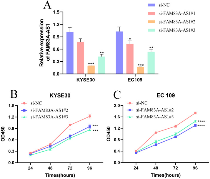
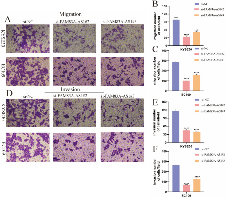
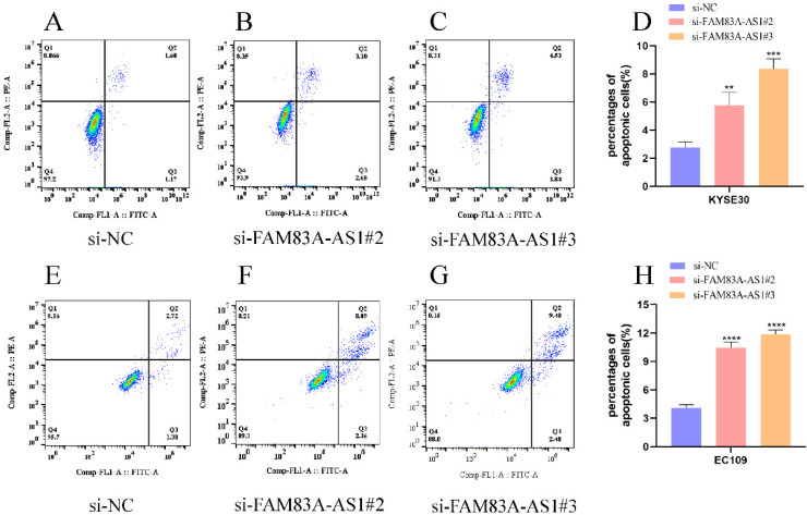
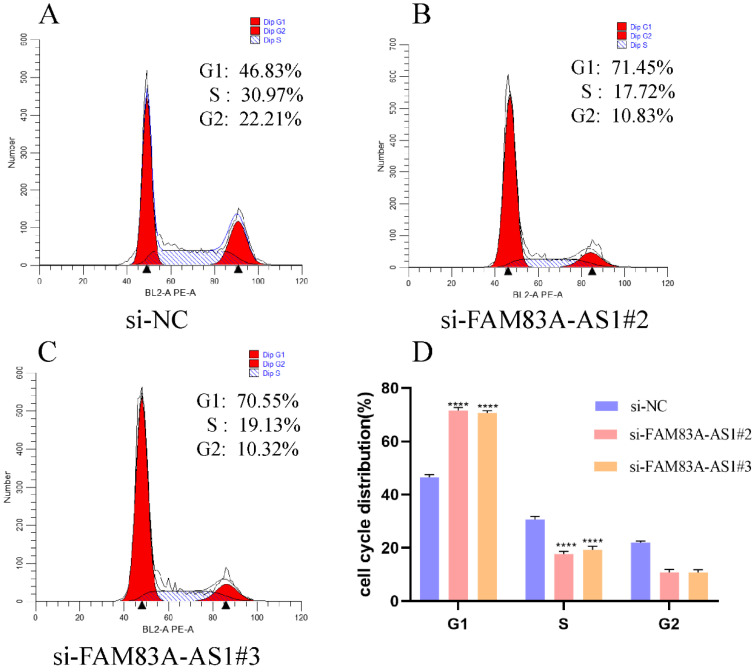
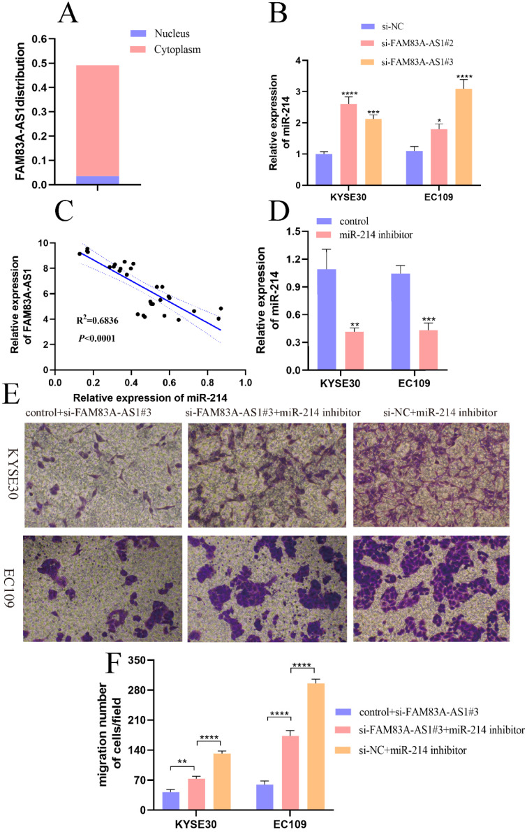
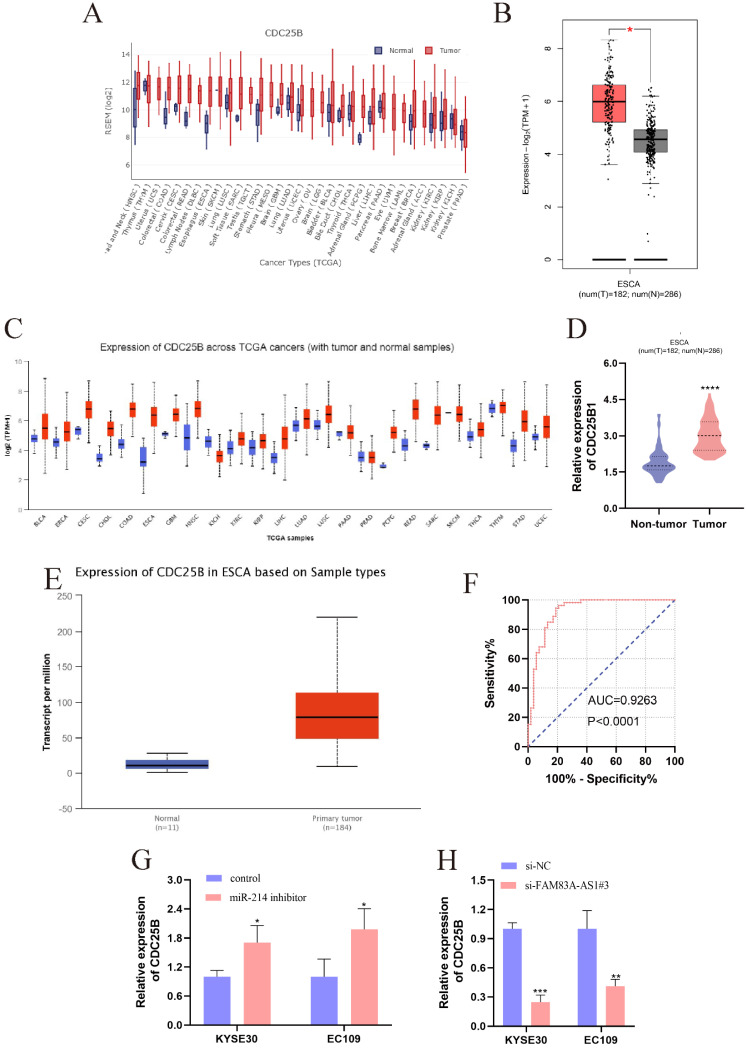
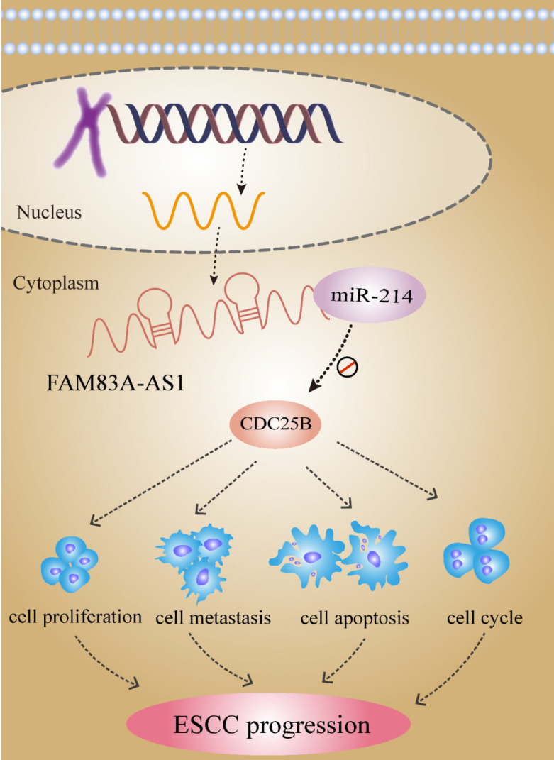
Background: Recent researches have pinpointed that long non-coding RNA (lncRNA) was tightly related to the carcinogenesis. However, the function of lncRNA in esophageal cell squamous carcinoma (ESCC) remains to be explored. In the current study, we assessed the expression pattern and the biological function of FAM83A-AS1 in ESCC. Methods: qRT-PCR was used to detect the expression of FAM83A-AS1, miR-214, and CDC25B expression in ESCC tissues and cell lines. CCK-8, transwell, apoptosis and cell cycle assays were performed to define the function of FAM83A-AS1 in ESCC cells. Furthermore, the regulation of miR-214 by FAM83A-AS1 was defined by qRT- PCR and rescue assays. In addition, the association between CDC25B, miR-214, CDC25B was confirmed by qRT-PCR. Results: Here, we discovered that FAM83A-AS1 was strongly expressed in ESCC tissues. FAM83A-AS1 abundance was associated with TNM stages and the differentiation grade of ESCC patients. The receiver operating characteristic curve (ROC) analysis indicated the high accuracy of FAM83A-AS1 in ESCC diagnosis. Functionally, inhibiting FAM83A-AS1 repressed cell proliferation, migration, and invasion in ESCC. In addition, we found that FAM83A-AS1 accelerated the cell cycle while inhibited cell apoptosis. Mechanistically, we found that FAM83A-AS1 regulated miR-214 expression, and there was a negative correlation between miR-214 and FAM83A-AS1 in ESCC. Rescue assay indicated that miR-214 could impair the suppressing effect of cell migration induced by FAM83A-AS1 depletion. Furthermore, CDC25B was a direct target of miR-214, and FAM83A-AS1 enhanced CDC25B expression while miR-214 positively CDC25B expression in ESCC. Conclusions: Collectively, we concluded that FAM83A-AS1 facilitated ESCC progression by regulating the miR-214/CDC25B axis. Our study showed FAM83A-AS1 may act as a promising target for ESCC diagnosis and therapy.

Keywords: CDC25B; ESCC; FAM83A-AS1; lncRNA; miR-214.


Conflict of interest statement

Competing Interests: The authors have declared that no competing interest exists.

Figures

Figure 1
Figure 1
FAM83A-AS1 was strongly overexpressed in ESCC tissues. (A-B) The bioinformatic analysis retrieved from GEPIA and lnCAR illuminated that FAM83A-AS1 was accumulated in ESCC tissues (ESCA indicates the esophageal carcinoma). (C) FAM83A-AS1 was significantly increased in 51 ESCC tissues compared with the adjacent tumor tissues. (D) ROC analysis showed a high accuracy of FAM83A-AS1 in ESCC diagnosis. (E) Kaplan-Meier analysis revealed the association between FAM83A-AS1 expression and survival time of ESCC patients. *P<0.05, ****P<0.0001.
Figure 2
Figure 2
Inhibiting FAM83A-AS1 impeded ESCC cell proliferation. (A) The treatment of si-FAM83A-AS1 in KYSE30 and EC109 cells hampered FAM83A-AS1 expression. (B-C) CCK-8 assay results demonstrated that FAM83A-AS1 promoted ESCC cell generation. *P<0.05, **P<0.01, ***P<0.001, ****P<0.0001.
Figure 3
Figure 3
FAM83A-AS1 increased ESCC cell metastasis. (A-C) FAM83A-AS1 depletion suppressed cell migration activity in KYSE30 and EC109 cells. (D-F) The invasive ability was significantly reduced after knocking down FAM83A-AS1 in KYSE30 and EC109 cells. ****P<0.0001.
Figure 4
Figure 4
FAM83A-AS1 negatively regulated cell apoptosis in ESCC. (A-D) Flow cell cytometry was used to examine the apoptosis events in ESCC cells with different treatments. The total number of apoptosis cells was growing after inhibiting FAM83A-AS1 in KYSE30 cell. (E-H) Flow cell cytometry analysis based on EC109 treated with si-FAM83A-AS1 produced the resembling results. *P<0.05, **P<0.01, ***P<0.001, ****P<0.0001.
Figure 5
Figure 5
FAM83A-AS1 regulated cell cycle distribution in ESCC. (A-D) EC109 cell cycle distribution with flow cell cytometry was tested according to the instructions of the cell cycle detection kit. There was a remarkable rise of cells in the G1 phase while the count of cells in the S phase was reduced. ***P<0.001, ****P<0.0001.
Figure 6
Figure 6
FAM83A-AS1 promoted ESCC development by regulating miR-214. (A) The subcellular localization of FAM83A-AS1 in the cell. (B) FAM83A-AS1 silencing produced an increase of miR-214 in ESCC cells. (C) The miR-214 expression pattern was negatively related to FAM83A-AS1 in ESCC tissues. (D) miR-214 inhibitor repressed miR-214 expression in ESCC cell. (E-F) The weakening of cell migration in ESCC cells induced by FAM83A-AS1 depletion could be reversed by miR-214 inhibitor partly. *P<0.05, **P<0.01, ***P<0.001, ****P<0.0001.
Figure 7
Figure 7
The high expression of CDC25B in ESCC and the regulations between CDC25B, miR-214, and FAM83A-AS1. (A) The pan-cancer analysis of CDC25B from GEDS. (B) CDC25B was significantly over-expressed in ESCC tissues, which was validated by GEPIA. (C) The pan-cancer analysis of CDC25B from Uclcan. (D-E) The up-regulation of CDC25B in ESCC was confirmed by oncomine and Uclcan. (F) The diagnostic value of CDC25B is based on the ROC analysis in ESCC. (G) miR-214 inhibitor engendered the augment of CDC25B in ESCC cells. (H) FAM83A-AS1 knockdown caused the reduction of CDC25B expression in ESCC cells. *P<0.05, **P<0.01, ***P<0.001, ****P<0.0001.
Figure 8
Figure 8
lncRNA FAM83A-AS1 dives ESCC progression by regulating the miR-214/CDC25B axis.

References

    1. Chen Y, Lu Y, Ren Y, Yuan J, Zhang N, Kimball H. et al. Starvation-induced suppression of DAZAP1 by miR-10b integrates splicing control into TSC2-regulated oncogenic autophagy in esophageal squamous cell carcinoma. Theranostics. 2020;10(11):4983–4996. - PMC - PubMed
    1. Teng H, Xue M, Liang J, Wang X, Wang L, Wei W. et al. Inter- and intratumor DNA methylation heterogeneity associated with lymph node metastasis and prognosis of esophageal squamous cell carcinoma. Theranostics. 2020;10(7):3035–3048. - PMC - PubMed
    1. Fong L, Taccioli C, Palamarchuk A, Tagliazucchi G, Jing R, Smalley K. et al. Abrogation of esophageal carcinoma development in miR-31 knockout rats. Proceedings of the National Academy of Sciences of the United States of America. 2020;117(11):6075–6085. - PMC - PubMed
    1. Liu K, Xie F, Zhao T, Zhang R, Gao A, Chen Y. et al. Targeting SOX2 Protein with Peptide Aptamers for Therapeutic Gains against Esophageal Squamous Cell Carcinoma. Molecular therapy: the journal of the American Society of Gene Therapy. 2020;28(3):901–913. - PMC - PubMed
    1. Sun K, Zhao X, Wan J, Yang L, Chu J, Dong S. et al. The diagnostic value of long non-coding RNA MIR31HG and its role in esophageal squamous cell carcinoma. Life Science. 2018;202:124–30. - PubMed