

Development of a Platelet Lysate-Based Printable, Transparent Biomaterial With Regenerative Potential for Epithelial Corneal Injuries
- PMID: 33442494
- PMCID: PMC7779874
- DOI: 10.1167/tvst.9.13.40
Development of a Platelet Lysate-Based Printable, Transparent Biomaterial With Regenerative Potential for Epithelial Corneal Injuries
Abstract
Purpose: To develop a human platelet lysate (hPL)-based bioink that is transparent and also encompasses the regenerative properties of hPL to facilitate wound healing.
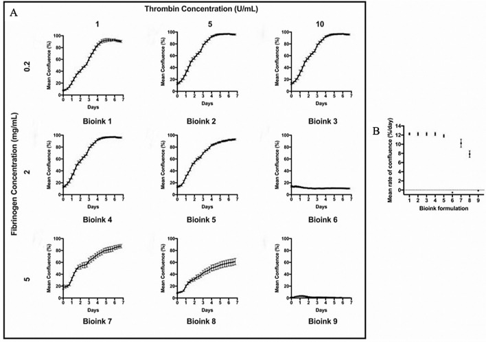
Methods: The effect of different batches of hPLand fetal bovine serum (FBS) on corneal epithelial cell growth and scratch assay was first examined using Incucyte Zoom. Various combinations of human fibrinogen (concentration range from 0.2 to 5 mg/mL) and thrombin (concentration from 1 to 10 U/mL) were combined with hPL to generate nine types of potential bioink. Rheology, transparency, and cell compatibility of bioinks were assessed and compared. The final selected bioink was used in an ex vivo corneal model to examine its ability in re-epithelization.
Results: No significant difference was detected in cell proliferation and wound healing tests between different hPL batches at the same concentration. Scratch assay data showed that hPL had significantly higher effect on wound healing than FBS. Comparing across the nine bioinks, bioink 5 consisting of 10% hPL, 2 mg/mL fibrinogen, and 5 U/mL thrombin demonstrated all required mechanical and cellular properties and was able to regenerate the full-thickness epithelium ex vivo.
Conclusions: The results showed that a transparent and adhesive bioink can be generated by combining hPL, fibrinogen, and thrombin together. The bioink can be directly applied to a human cornea to promote corneal re-epithelization with huge potential applications in corneal injuries.
Translational relevance: The developed transparent hPL-based ink with its adhesive and healing ability showed that it could be used as a new treatment option for corneal injuries.
Keywords: bioink; corneal wound healing; fibrin; human platelet lysate.
Copyright 2020 The Authors.
Conflict of interest statement
Disclosure: H. Frazer (P); J. You, iFix Medical (P, I, F); Z. Chen, None; S. Sayyar, None; X. Liu, None; A. Taylor, None; C. Hodge, None; G. Wallace, None; G. Sutton, iFix Medical (P, I)
Figures


References
-
- Flaxman SR, Bourne RRA, Resnikoff S, et al.. Global causes of blindness and distance vision impairment 1990–2020: a systematic review and meta-analysis. Lancet Glob Health. 2017; 5: e1221–e1234. - PubMed
-
- McCarty CA, Fu CL, Taylor HR. Epidemiology of ocular trauma in Australia. Ophthalmology. 1999; 106: 1847–1852. - PubMed
-
- Ashby BD, Garrett Q, Willcox MD. Corneal injuries and wound healing—review of processes and therapies. Austin J Clin Ophthalmol. 2014; 1: 1–25.
Publication types
MeSH terms
Substances
LinkOut - more resources
Full Text Sources
Medical
Research Materials
