Clinico-Radio-Pathological and Molecular Features of Hepatocellular Carcinomas with Keratin 19 Expression
- PMID: 33442539
- PMCID: PMC7768132
- DOI: 10.1159/000510522
Clinico-Radio-Pathological and Molecular Features of Hepatocellular Carcinomas with Keratin 19 Expression
Abstract
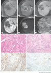
Hepatocellular carcinoma (HCC) is a heterogeneous neoplasm, both from the molecular and histomorphological aspects. One example of heterogeneity is the expression of keratin 19 (K19) in a subset (4-28%) of HCCs. The presence of K19 expression in HCCs has important clinical implications, as K19-positive HCCs have been associated with aggressive tumor biology and poor prognosis. Histomorphologically, K19-positive HCCs demonstrate a more infiltrative appearance, poor histological differentiation, more frequent vascular invasion, and more intratumoral fibrous stroma than K19-negative conventional HCCs. From the molecular aspect, K19-positive HCCs have been matched with various gene signatures that have been associated with stemness and poor prognosis, including the G1-3 groups, S2 class, cluster A, proliferation signature, and vascular invasion signature. K19-positive HCCs also show upregulated signatures related to transforming growth factor-β pathway and epithelial-to-mesenchymal transition. The main regulators of K19 expression include hepatocyte growth factor-MET paracrine signaling by cancer-associated fibroblast, epidermal growth factor-epidermal growth factor receptor signaling, laminin, and DNA methylation. Clinically, higher serum alpha-fetoprotein levels, frequent association with chronic hepatitis B, more invasive growth, and lymph node metastasis have been shown to be characteristics of K19-positive HCCs. Radiologic features including atypical enhancement patterns, absence of tumor capsules, and irregular tumor margins can be a clue for K19-positive HCCs. From a therapeutic standpoint, K19-positive HCCs have been associated with poor outcomes after curative resection or liver transplantation, and resistance to systemic chemotherapy and locoregional treatment, including transarterial chemoembolization and radiofrequency ablation. In this review, we summarize the currently available knowledge on the clinico-radio-pathological and molecular features of K19-expressing HCCs, including a detailed discussion on the regulation mechanism of these tumors.
Keywords: Hepatocellular carcinoma; Invasiveness; Keratin 19; Poor prognosis; Treatment resistance.
Copyright © 2020 by S. Karger AG, Basel.
Conflict of interest statement
The authors have no conflicts of interest to declare.
Figures


References
-
- Bray F, Ferlay J, Soerjomataram I, Siegel RL, Torre LA, Jemal A. Global cancer statistics 2018: GLOBOCAN estimates of incidence and mortality worldwide for 36 cancers in 185 countries. CA Cancer J Clin. 2018;68((6)):394–424. - PubMed
-
- Torbenson MS, Ng IOL, Park YN, Roncalli M, Sakamoto M. Hepatocellular carcinoma. In: WHO Classification of Tumours Editorial Board, editor. Digestive system tumours. Lyon: International Agency for Research on Cancer; 2019. pp. p.229–39.
-
- Roskams T. Liver stem cells and their implication in hepatocellular and cholangiocarcinoma. Oncogene. 2006;25((27)):3818–22. - PubMed
-
- Durnez A, Verslype C, Nevens F, Fevery J, Aerts R, Pirenne J, et al. The clinicopathological and prognostic relevance of cytokeratin 7 and 19 expression in hepatocellular carcinoma. A possible progenitor cell origin. Histopathology. 2006;49((2)):138–51. - PubMed
Publication types
LinkOut - more resources
Full Text Sources
Research Materials
Miscellaneous

