[Preprint]. 2021 Mar 18:2021.01.08.425793.
doi: 10.1101/2021.01.08.425793.

Extracellular vimentin as a target against SARS-CoV-2 host cell invasion

Affiliations

Extracellular vimentin as a target against SARS-CoV-2 host cell invasion

Łukasz Suprewicz et al. bioRxiv. .

Update in

  • Extracellular Vimentin as a Target Against SARS-CoV-2 Host Cell Invasion.
    Suprewicz Ł, Swoger M, Gupta S, Piktel E, Byfield FJ, Iwamoto DV, Germann D, Reszeć J, Marcińczyk N, Carroll RJ, Janmey PA, Schwarz JM, Bucki R, Patteson AE. Suprewicz Ł, et al. Small. 2022 Feb;18(6):e2105640. doi: 10.1002/smll.202105640. Epub 2021 Dec 5. Small. 2022. PMID: 34866333 Free PMC article.

Abstract

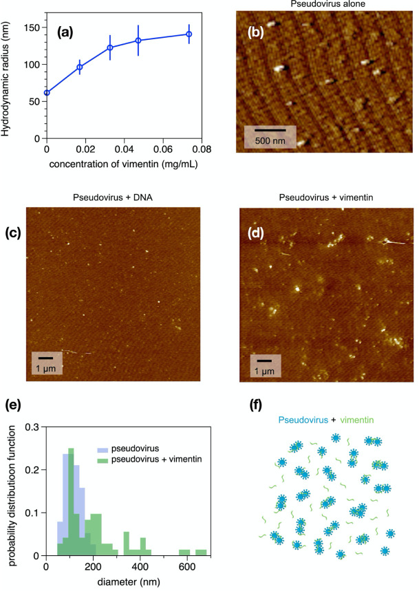
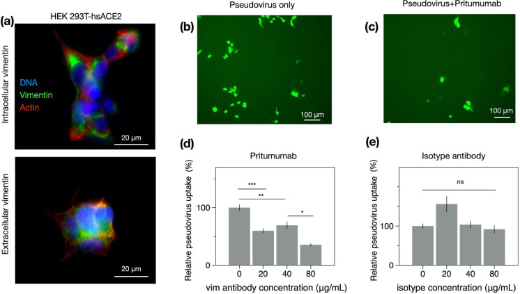
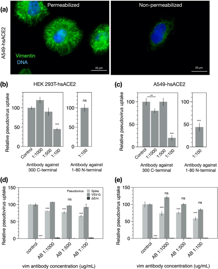
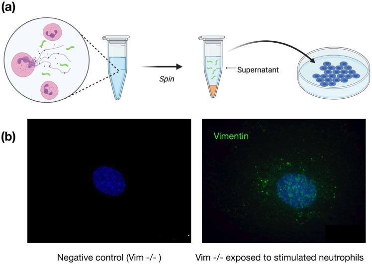
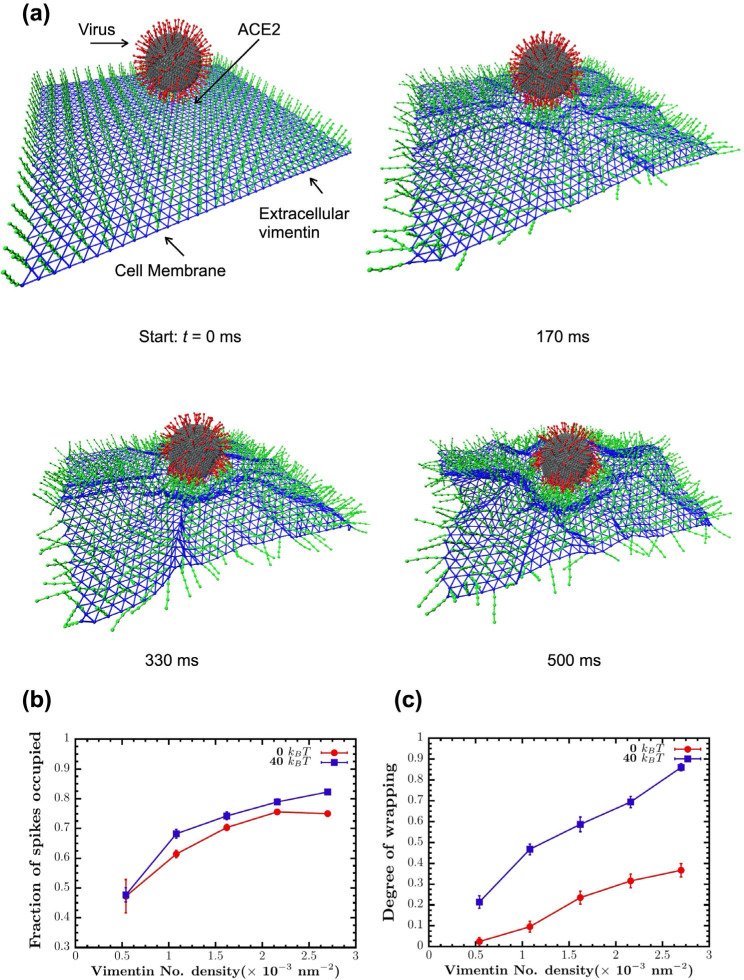
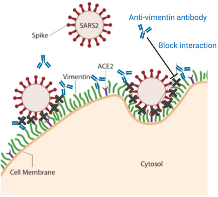
Infection of human cells by pathogens, including SARS-CoV-2, typically proceeds by cell surface binding to a crucial receptor. In the case of SARS-CoV-2, angiotensin-converting enzyme 2 (ACE2) has been identified as a necessary receptor, but not all ACE2-expressing cells are equally infected, suggesting that other extracellular factors are involved in host cell invasion by SARS-CoV-2. Vimentin is an intermediate filament protein that is increasingly recognized as being present on the extracellular surface of a subset of cell types, where it can bind to and facilitate pathogens' cellular uptake. Here, we present evidence that extracellular vimentin might act as a critical component of the SARS-CoV-2 spike protein-ACE2 complex in mediating SARS-CoV-2 cell entry. We demonstrate direct binding between vimentin and SARS-CoV-2 pseudovirus coated with the SARS-CoV-2 spike protein and show that antibodies against vimentin block in vitro SARS-CoV-2 pseudovirus infection of ACE2-expressing cells. Our results suggest new therapeutic strategies for preventing and slowing SARS-CoV-2 infection, focusing on targeting cell host surface vimentin.


Conflict of interest statement

Competing Interests: The authors declare no competing interests.

Figures

Figure 1.
Figure 1.. Presence of extracellular vimentin in human lung, airway fluids, and fat tissue,
(a) Positive staining for extracellular vimentin (green) in human lung, fat tissue, and sputum obtained from cystic fibrosis (CF) patients. Vimentin appears on the apical side of type I and type II pneumocytes. DNA stained with DAPI. (b) There are numerous internal and exogenous pathways by which vimentin may be found in lung epithelia and other tissues, in either intracellular or cell surface forms (shown as green filaments). Vimentin is expressed directly by mesenchymal cells, cells having undergone EMT, cancer cells, senescent fibroblasts, and interestingly by cells bound and infected by the SARS-CoV virus (see Table 1). Exogenous sources of vimentin are largely related to immune response and tissue injury in the form of vimentin exported by neutrophils, T-lymphocytes, monocytes/macrophages, and exosomes. Schematics generated with Biorender.com.
Figure 2.
Figure 2.. Binding of vimentin to SARS-CoV-2 pseudovirus.
Purified human recombinant vimentin was added to suspensions of SARS-CoV-2 spike protein-containing pseudovirus, and their size was measured by dynamic light scattering (a) and atomic force microscopy (b-e). The size of the pseudovirus increased from 60 nm to 150 nm after addition of 0.07 mg/ml vimentin (panel a). Panel b-d: The pseudoviruses were imaged using atomic force microscopy before and after addition of either DNA (0.08 mg/mL) or vimentin (0.07 mg/ml). The probability distribution functions (e) show that the average size of pseudovirus imaged by AFM confirms the change in size detected by DLS. (f) Schematic representation indicating how binding of vimentin to SARS-CoV-2 spike protein could couple particles together and increase their effective radii.
Figure 3.
Figure 3.. Anti-vimentin antibodies block uptake of SARS-CoV-2 pseudovirus in HEK 293T-hsACE2.
(a) Positive staining for extracellular vimentin in mouse embryonic fibroblasts and human kidney epithelial cells HEK 293T-hs ACE2 and mouse embryonic fibroblasts. Images show vimentin (green), actin (red), and DNA (blue). To stain for extracellular vimentin, cells were exposed to a primary polyclonal chicken anti-vimentin antibody, before fixation, then permeabilized and stained for actin. (b) Schematic of experimental design of pseudovirus infection studies. HEK cells are pre-exposed to the anti-vimentin antibody Pritumumab before infection by pseudovirus bearing the SARS-CoV-2 spike protein and a GFP reporter, (c) Representative fluorescence images showing cells expressing GFP after pseudovirus exposure with and without Pritumumab treatment, (d) Pritumumab inhibits cellular infection by up to 60%. (e) Use of an isotype antibody does not inhibit infection, corroborating a specific interaction between the SARS-CoV-2 spike protein and extracellular vimentin. (f) Use of another vimentin antibody, a chicken IgY polyclonal antibody, also blocks cellular transduction by SARS-CoV-2 pseudovirus. Denotations: *, P ≤ 0.05; **, P < 0.01; ***, P < 0.001; NS, P > 0.05.
Figure 4.
Figure 4.. SARS-CoV-2 pseudovirus transduction results based on plate readers for HEK 293T-hsACE2 and A549-hsACE2 cells,
(a) Extracellular vimentin was detected on A549-hsACE2 as shown by immunofluorescence images, (b) The recombinant Anti-Vimentin antibody - rabbit monoclonal IgG, which binds to the vimentin C-terminus blocks uptake in both HEK 293T-hsACE2 and (c) A549-hsACE2 cells. The primary anti-vimentin polyclonal rabbit antibody that targets the phosphorylation site of SER56 on the N-terminus blocks uptake in A549-hsACE2 cells but not HEK 293T-hsACE2 cells. Denotations: *, P ≤ 0.05; **, P < 0.01; ***, P < 0.001; NS, P > 0.05.
Figure 5.
Figure 5.. Acquisition of cell surface vimentin from extracellular environment,
(a) Neutrophil NETosis stimulates disassembly of the vimentin network and enables its release into the extracellular space, (b) Immunofluorescence images of vimentin-null mEF staining positive for extracellular vimentin after exposure to supernatant of NETosis-activated neutrophils, indicating the acquisition of extracellular vimentin by cells that do not express vimentin. Schematics generated with Biorender.com.
Figure 6.
Figure 6.. Vimentin’s involvement in spike 2-ACE2 interactions.
(a) Cell surface vimentin acts as a co-receptor that enhances binding to the SARS-CoV-2 virus in either direct fusion or endocytic pathways to enhance wrapping and endocytosis of the virus. A molecular dynamics simulation of the SARS-CoV-2 virus at an elastic shell membrane shows that binding of extracellular vimentin with the virus spike protein facilitates wrapping of cell membrane around the virus. Both the fraction of spikes bound to surface vimentin (b) and the degree of membrane wrapping (c) increases as the number density of surface vimentin increases. Finite membrane bending rigidity (40 kB T, blue squares) enhances wrapping compared to the case without bending rigidity (0 kB T, red circles).
Figure 7.
Figure 7.. Extracellular vimentin as a potential target for inhibiting SARS-CoV-2 entry.
A diagram of proposed mechanism of action. Here, cell surface vimentin (green) acts as a co-receptor that binds to SARS-CoV-2 spike protein. Blocking this interaction via the anti-vimentin antibody Pritumumab reduces cell surface binding of the virus and cellular infection. Schematics generated with Biorender.com.

Similar articles

Cited by

References

    1. Wrapp D. et al., Cryo-EM structure of the 2019-nCoV spike in the prefusion conformation. Science 367,1260–1263 (2020). - PMC - PubMed
    1. Walls A. C. et al., Structure, function, and antigenicity of the SARS-CoV-2 spike glycoprotein. Cell, (2020). - PMC - PubMed
    1. Synowiec A., Szczepański A., Barreto-Duran E., Lie L. K., Pyre K., Severe Acute Respiratory Syndrome Coronavirus 2 (SARS-CoV-2): a Systemic Infection. Clinical Microbiology Reviews 34, e00133–00120 (2021). - PMC - PubMed
    1. Hikmet F. et al., The protein expression profile of ACE2 in human tissues. Mol Syst Biol 16, e9610 (2020). - PMC - PubMed
    1. Aguiar J. A. et al., Gene expression and in situ protein profiling of candidate SARS-CoV-2 receptors in human airway epithelial cells and lung tissue. European Respiratory Journal 56, 2001123 (2020). - PMC - PubMed

Publication types

LinkOut - more resources