[Preprint]. 2021 Jan 19:2021.01.06.425627.
doi: 10.1101/2021.01.06.425627.

Neuropilin-1 Assists SARS-CoV-2 Infection by Stimulating the Separation of Spike Protein Domains S1 and S2

Affiliations

Neuropilin-1 Assists SARS-CoV-2 Infection by Stimulating the Separation of Spike Protein Domains S1 and S2

Zhen-Lu Li et al. bioRxiv. .

Update in

Abstract

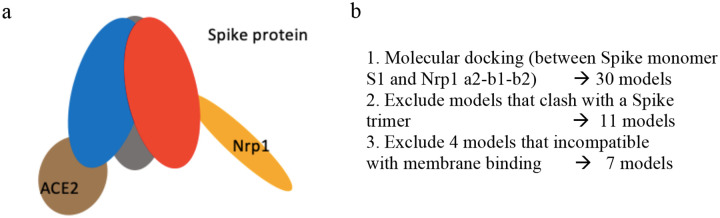
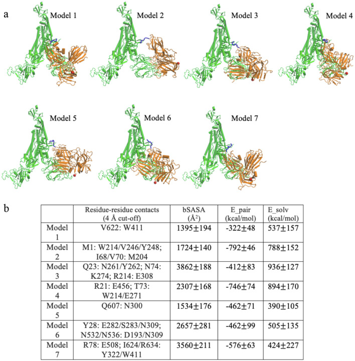
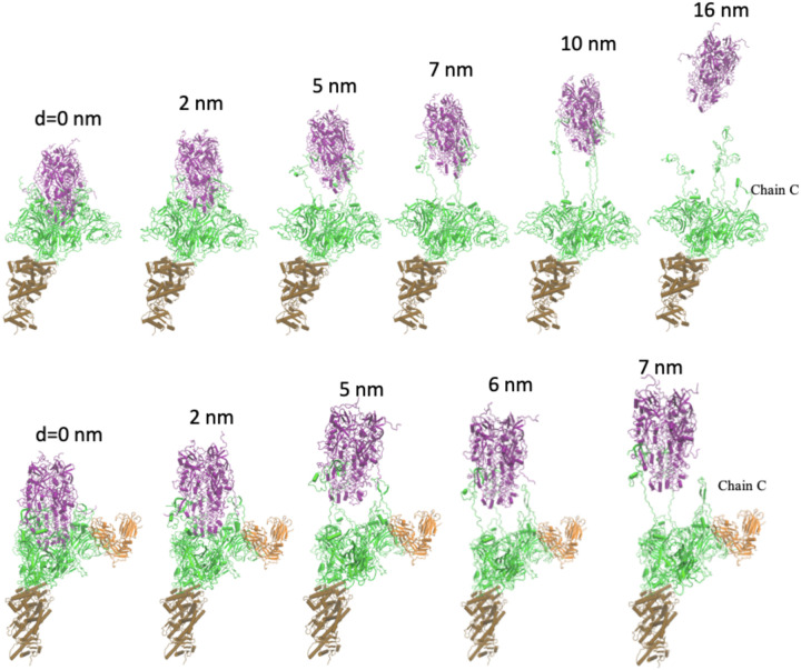
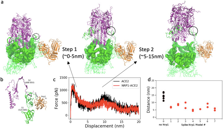
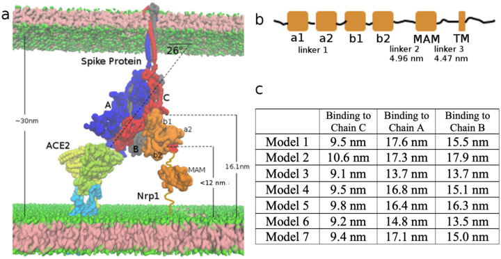
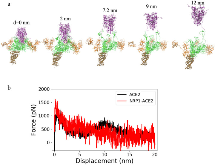
The cell surface receptor Neuropilin-1 (Nrp1) was recently identified as a host factor for SARS-CoV-2 entry. As the Spike protein of SARS-CoV-2 is cleaved into the S1 and the S2 domain by furin protease, Nrp1 binds to the newly created C-terminal RRAR amino acid sequence of the S1 domain. In this study, we model the association of a Nrp1 (a2-b1-b2) protein with the Spike protein computationally and analyze the topological constraints in the SARS-CoV-2 Spike protein for binding with Nrp1 and ACE2. Importantly, we study the exit mechanism of S2 from the S1 domain with the assistance of ACE2 as well as Nrp1 by molecular dynamics pulling simulations. In the presence of Nrp1, by binding the S1 more strongly to the host membrane, there is a high probability of S2 being pulled out, rather than S1 being stretched. Thus, Nrp1 binding could stimulate the exit of S2 from the S1 domain, which will likely increase virus infectivity as the liberated S2 domain mediates the fusion of virus and host membranes. Understanding of such a Nrp1-assisted viral infection opens the gate for the generation of protein-protein inhibitors, such as antibodies, which could attenuate the infection mechanism and protect certain cells in a future combination therapy.


Conflict of interest statement

Competing Interests The authors declare no competing interests.

Figures

Figure 1:
Figure 1:
(a) Schematic Model of Spike Protein trimer bound to one ACE2 and one Nrp1 receptor. (b) Molecular docking yields 7 possible binding models which satisfy geometric constraints.
Figure 2:
Figure 2:
Binding modes for 7 predicted models. (a) Models, shown for Chain C: Nrp1 a2-b1-b2 binding. The TNSPRRAR motif is marked in blue. The last residue of Nrp1 a2-b1-b2 domain is marked in red. (b) Noticeable residue-residue contacts (< 0.4 nm) between Spike protein and Nrp1 in different models (except the RRAR binding region). Together with residue-residue contacts (< 0.6 nm) the proteins are seen to interact via domains a2,b1 and b2 on Nrp1 and a limited number of regions N1-N4, RBDN, RBDC, C1-C3, the very C-term. of S1 and beginning of S2 (see Table S1 for details). Buried SASA, pair interaction energy and changes of solvation energy for different models. The interaction energy (based upon pairwise vdW and electrostatic forces) are calculated between Nrp1 a2-b1-b2 with a full Spike trimer (Chain A, B, C). The models shown are listed in order of their population near cluster centers (models) 1 to 7 (most to least).
Figure 3:
Figure 3:
Separation process of S2 and S1 domain of Chain C with/without Nrp1; here shown in absence of Nrp1, top (1st. of 7 simulations shown) and for Nrp1: Spike model 7, bottom. (Color coding: S1 in green and S2 in purple; Nrp1 in orange) The displacement of S2 domain center of mass relative to initial position is given as distance d. The time interval between the structures is 2, 5, 7, 10, 16 ns or 2, 5, 6, 7 ns in the pulling simulation (1 nm/ns).
Figure 4:
Figure 4:
(a) Two steps of S2 exit from S1 domain: S1 regions that cap S2 domain (purple) are highlighted in surface representation in green. (b) S1:S2 contacts at the cleavage site at the start of simulation (model 7 at 0 ns). (c) Pulling force versus displacement of S2 domain (with and without Nrp1). Averaged over simulations of 7 models for simulations without and with Nrp1. (d) Distance between S1 and S2 centers of mass of Chain C of the Spike protein at the time of separation (nearest distance >5Å) between the two domains (of chain C). Two repeats were done for each model except model 5, where Nrp1:S1 binding is easily disconnected during the pulling of S2 domain.
Figure 5:
Figure 5:
Topological constraints of Nrp1 on binding with Spike protein trimer, bound to an ACE2 dimer. (a) ACE2 (yellow and limone, which also go into and through the membrane) and B0AT1(light blue) 2:2 dimer complex at the bottom left. Spike protein trimer (Chain A, B, C in blue, grey and red) in the middle. Nrp1 (orange) at the bottom right of the figure. (b) Linear domain arrangement in the sequence of Nrp1 receptor. (c) Estimated distance of C-terminus of the b2 domain to the membrane bilayer lipid headgroups in the different models with Nrp1 bound to Chain A, B, C.
Figure 6:
Figure 6:
(a) Separation process of S2 and S1 domain of Chain A, B and C with three Nrp1s binding to each unit of the Spike protein trimer. The displacement of the S2 domain relative to initial position is shown. (b) Pulling force versus displacement of S2 domain with three Nrp1 binding. Averaged over 2 simulations for model 1 (red) whereas the ACE2 data is taken from Fig. 4c.

References

    1. Daly JL, Simonetti B., An ton-Plagaro C, et al., Neuropilin-1 is a host factor for SARS-CoV-2 infection. Science 2020, 370(6518):861–865. - PMC - PubMed
    1. Cantuti-Castelvetri L, Ojha R, Pedro LD, et al., Neuropilin-1 facilitates SARS-CoV-2 cell entry and infectivity. Science 2020, 370(6518):856–860. - PMC - PubMed
    1. Moutal A, Martin LF, Boinon L, Gomez K, et al., SARS-CoV-2 Spike protein hijacks VEGFA/Neuropilin-1 receptor signaling to induce analgesa. bioRxiv 2020. 10.1101/2020.01.17.209288. - DOI - PubMed
    1. Hoffmann M, et al. SARS-CoV-2 cell entry depends on ACE2 and TMPRSS2 and is blocked by a clinically proven protease inhibitor. Cell 2020; 181, 271–280. - PMC - PubMed
    1. Shang J, et al. Cell entry mechanisms of SARS-CoV-2. Proc. Natl. Acad. Sci. USA 2020;117,11727–11734. - PMC - PubMed

Publication types