. 2021 May 14;22(10):1841-1851.
doi: 10.1002/cbic.202000870. Epub 2021 Feb 18.

SP3-FAIMS Chemoproteomics for High-Coverage Profiling of the Human Cysteinome*

Affiliations

SP3-FAIMS Chemoproteomics for High-Coverage Profiling of the Human Cysteinome*

Tianyang Yan et al. Chembiochem. .

Abstract

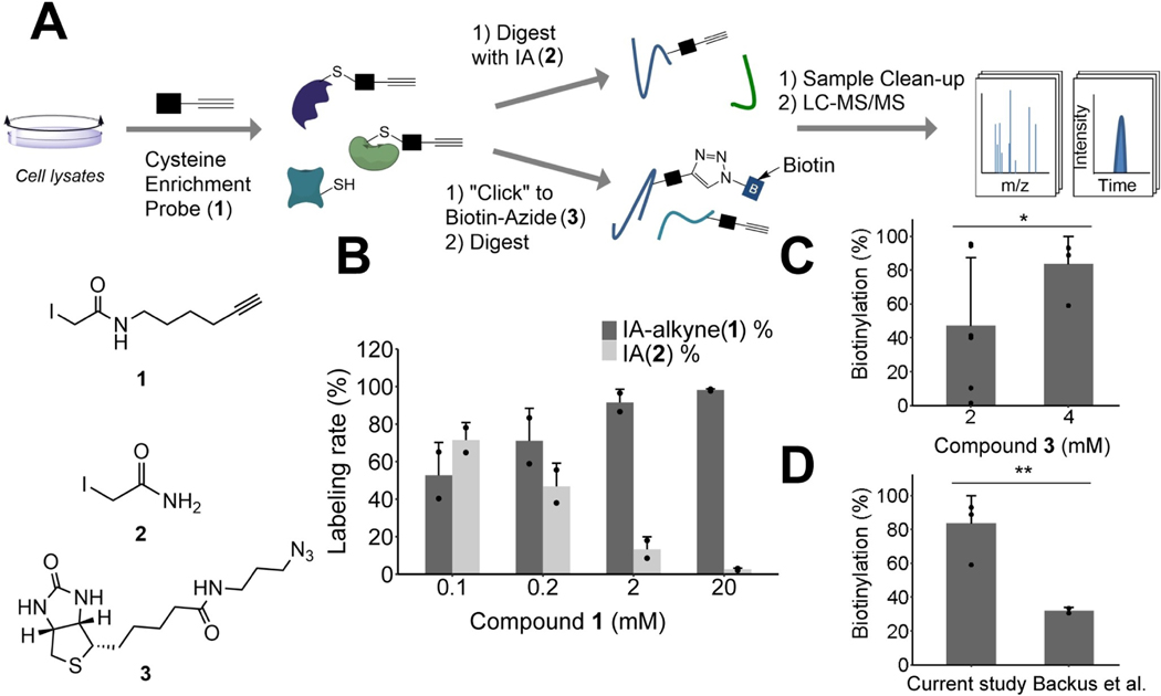
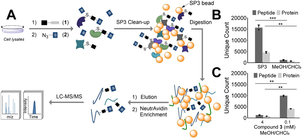
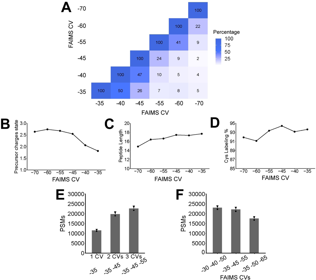
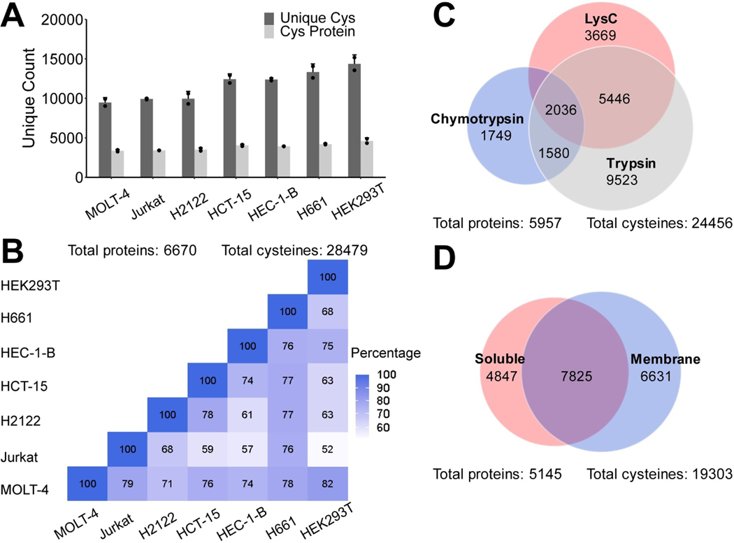
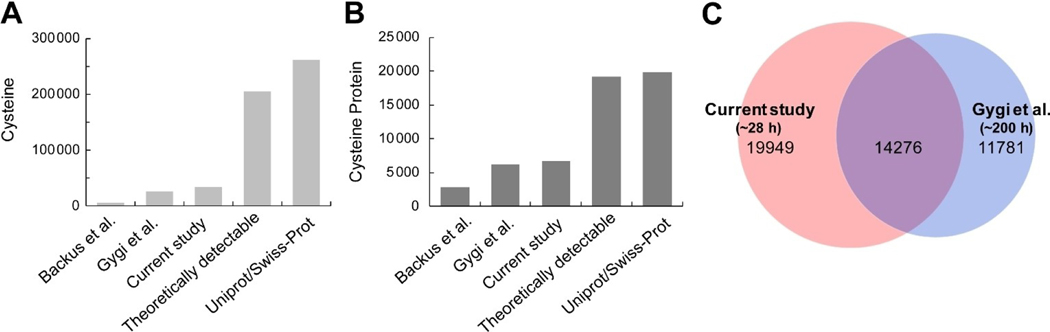
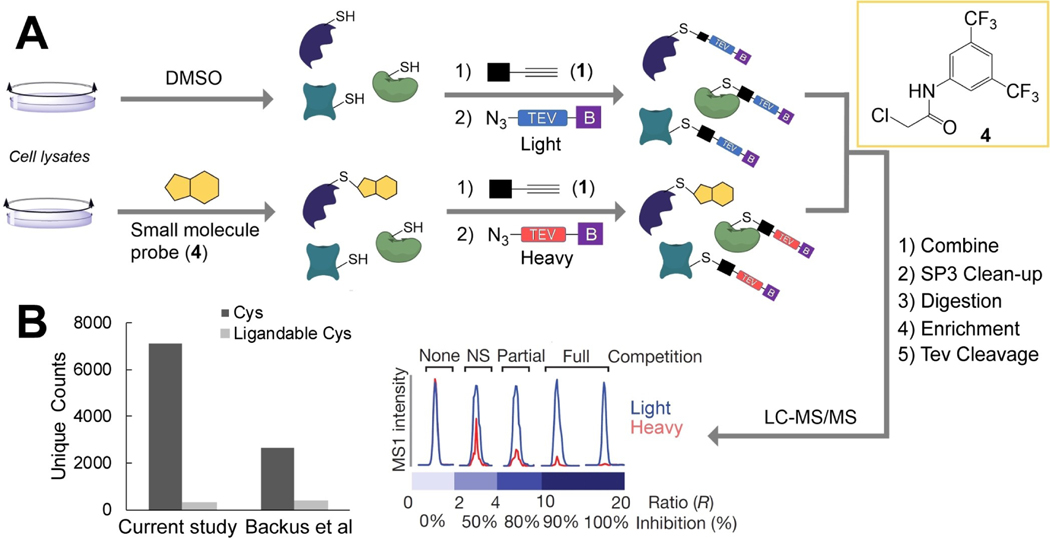
Chemoproteomics has enabled the rapid and proteome-wide discovery of functional, redox-sensitive, and ligandable cysteine residues. Despite widespread adoption and considerable advances in both sample-preparation workflows and MS instrumentation, chemoproteomics experiments still typically only identify a small fraction of all cysteines encoded by the human genome. Here, we develop an optimized sample-preparation workflow that combines enhanced peptide labeling with single-pot, solid-phase-enhanced sample-preparation (SP3) to improve the recovery of biotinylated peptides, even from small sample sizes. By combining this improved workflow with on-line high-field asymmetric waveform ion mobility spectrometry (FAIMS) separation of labeled peptides, we achieve unprecedented coverage of >14000 unique cysteines in a single-shot 70 min experiment. Showcasing the wide utility of the SP3-FAIMS chemoproteomic method, we find that it is also compatible with competitive small-molecule screening by isotopic tandem orthogonal proteolysis-activity-based protein profiling (isoTOP-ABPP). In aggregate, our analysis of 18 samples from seven cell lines identified 34225 unique cysteines using only ∼28 h of instrument time. The comprehensive spectral library and improved coverage provided by the SP3-FAIMS chemoproteomics method will provide the technical foundation for future studies aimed at deciphering the functions and druggability of the human cysteineome.

Keywords: LC-MS/MS; chemoproteomics; cysteine; high-field asymmetric waveform ion mobility spectrometry (FAIMS); single-pot, solid-phase-enhanced sample-preparation (SP3).


Conflict of interest statement

Conflict of Interest

The authors declare no conflict of interest.

Figures

Figure 1.
Figure 1.
Proteomic analysis of cysteine-labeling efficiency. A) Workflow employed to assess IAA labeling (top) and CuAAC biotinylation (bottom) of cysteines.B) Percentage of cysteines labeled by 1 or 2, when denatured and DTT-reduced HEK239T lysates were treated with either 0.1, 0.2, 2, or 20 mM 1 followed by 20 mM 2. Experiments were performed in duplicate. C) Cysteine biotinylation rate for cell lysates treated with 2 mM 1 followed by CuAAC conjugation to either 2 or 4 mM 3. Experiments were performed with six replicates for 2 mM 3 and four replicates for 4 mM 3. D) Comparison of cysteine biotinylation rates for cell lysates treated with 2 mM 1 and 4 mM 3, as in (C), to 100 μM 1 and 100 μM 3, conditions that model those employed by prior studies.[12] Experiments were performed in duplicate. For (C) and (D), statistical significance was calculated with unpaired Student’s t-tests, *P<0.05, **P<0.005. All data can be found in Table S1.
Figure 2.
Figure 2.
Cysteine enrichment with single-pot, solid-phase-enhanced sample preparation (SP3). A) The schematic workflow of cysteine enrichment with SP3. B) Total number of unique proteins and cysteines identified with SP3 or MeOH/CHCl3 protein clean-up for samples treated with 2 mM IAA and 4 mM biotinazide. C) Comparison of unique proteins and cysteine peptides identified with MeOH/CHCl3 protein precipitation treated with 0.1 or 4 mM 3. For (B) and (C), statistical significance was calculated with unpaired Student’s t-tests, *P<0.05, **P<0.005, ***P<0.001. All experiments were performed in duplicate. All data can be found in Table S2.
Figure 3.
Figure 3.
Characterization of different FAIMS CV settings for cysteine enrichment. A) Similarity comparison of cysteine peptides identified with six different CVs. Distributions of B) precursor charge states, C) peptide length, and D) cysteine biotinylation rate across CV settings from −35 to−70 V. E) PSMs identified with different numbers of CVs, one CV (−35), two CVs (−35 and−45) and three CVs (−35, −45, and −55). F) PSMs identified with different three CV settings. All experiments were performed in duplicate. All data can be found in Table S3.
Figure 4.
Figure 4.
SP3-based cysteine profiles of different cell lines, with different proteases and fractionations. A) Total number of unique cysteines and cysteine proteins identified with SP3 in MOLT-4, HEC-1-B, HEK293T, H2122, HCT-15, Jurkat and H661 cell lines. B) Similarity comparison of cysteines identified in different cell lines. C) Venn diagram of unique cysteines identified with different proteases (trypsin, lysC and chymotrypsin) in HEK293T cells. D) Venn diagram of unique cysteines identified with soluble and membrane fractions of HEK293T cells. All experiments were performed in duplicate. All data can be found in Table S4.
Figure 5.
Figure 5.
A) Cysteines and B) cysteine proteins identified experimentally in Backus et al.,[12] Gygi et al.,[58] in the current study, those theoretically detectable based on in silico trypsin digestion, and all found in Uniprot/Swiss-Prot sequences. C) Venn diagram of unique cysteines identified in current study and Gygi et al. All data can be found in Table S5.
Figure 6.
Figure 6.
Chemoproteomic analysis of ligandable cysteines with SP3. A) Schematic workflow of competitive isoTOP-ABPP with SP3. B) Comparison of ligandable cysteines and total cysteines identified with isoTOP-ABPP treated with compound 4 in Backus et al.[12] and the current study. Experiments were performed in quadruplicate. All data can be found in Table S6.

References

    1. Drewes G, Knapp S, Trends Biotechnol. 2018, 36, 1275–1286. - PubMed
    1. Simon GM, Cravatt BF, J. Biol. Chem. 2010, 285, 11051–11055. - PMC - PubMed
    1. Shenoy VM, Thompson BR, Shi J, Zhu HJ, Smith DE, Amidon GL, Mol. Pharm. 2020, 17, 1706–1714. - PubMed
    1. Li W, Blankman JL, Cravatt BF, J. Am. Chem. Soc. 2007, 129, 9594–9595. - PubMed
    1. Hacker SM, Backus KM, Lazear MR, Forli S, Correia BE, Cravatt BF, Nat. Chem. 2017, 9, 1181–1190. - PMC - PubMed

Publication types