Review
. 2020 Dec;7(2):e001424.
doi: 10.1136/openhrt-2020-001424.

Manipulation of ACE2 expression in COVID-19

Affiliations
Review

Manipulation of ACE2 expression in COVID-19

Farhan Chaudhry et al. Open Heart. 2020 Dec.

Abstract

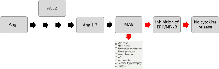
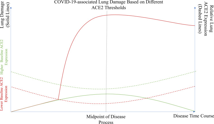
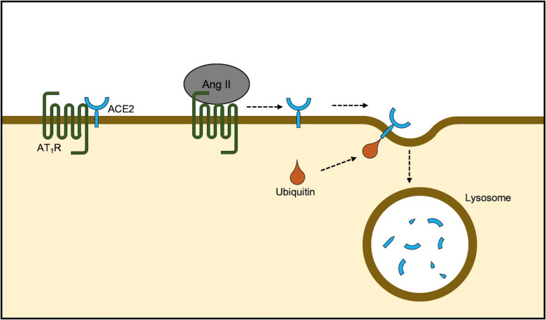
SARS-CoV-2 is the virus responsible for the ongoing COVID-19 outbreak. The virus uses ACE2 receptor for viral entry. ACE2 is part of the counter-regulatory renin-angiotensin-aldosterone system and is also expressed in the lower respiratory tract along the alveolar epithelium. There is, however, significant controversy regarding the role of ACE2 expression in COVID-19 pathogenesis. Some have argued that decreasing ACE2 expression would result in decreased susceptibility to the virus by decreasing available binding sites for SARS-CoV-2 and restricting viral entry into the cells. Others have argued that, like the pathogenesis of other viral pneumonias, including those stemming from previous severe acute respiratory syndrome (SARS) viruses, once SARS-CoV-2 binds to ACE2, it downregulates ACE2 expression. Lack of the favourable effects of ACE2 might exaggerate lung injury by a variety of mechanisms. In order to help address this controversy, we conducted a literature search and review of relevant preclinical and clinical publications pertaining to SARS-CoV-2, COVID-19, ACE2, viral pneumonia, SARS, acute respiratory distress syndrome and lung injury. Our review suggests, although controversial, that patients at increased susceptibility to COVID-19 complications may have reduced baseline ACE2, and by modulating ACE2 expression one can possibly improve COVID-19 outcomes. Herein, we elucidate why and how this potential mechanism might work.

Keywords: hypertension; intensive care; lung injury.


Conflict of interest statement

Competing interests: None declared.

Figures

Figure 1
Figure 1
SARS-CoV-2 entry into the cell. SARS-CoV-2 binds to ACE2 through the receptor-binding domain (RBD) of spike protein. The L455, F486, Q493, S494, N501 and Y505 residues in SARS-CoV-2 RBD interact with K31, E35, D38, M82 and K353 residues of ACE2 receptor. Notably, the RBD of SARS-CoV-2 tends to generate a large binding interface of 1204 Å, along with an extended contact with the N-terminal helix of ACE2, and the higher affinity of the virus for ACE2 binding. For entry, S1 and S2 subunits dissociate from each other through cleavage by a protease, such as TMPRSS2. Furin also cleaves the site between both S1 and S2 subunits of the SARS-CoV-2 spike protein and serves as the alternative pathway of activation, especially in cells with lower TMPRSS2 expression.
Figure 2
Figure 2
The ACE2 mechanism. Ang II is converted by ACE2 to become Ang-(1–7). Ang-(1–7) activates the MAS receptor signalling pathway, which then inhibits ERK/NF-κB, resulting in no cytokine release. Ang II, angiotensin II; ERK, extracellular signal-regulated kinase; MAS, mitochondrial assembly; NF-κB, nuclear factor kappa B; NO, nitric oxide; PSNS, parasympathetic nervous system; SNS, sympathetic nervous system.
Figure 3
Figure 3
SARS-CoV-2-associated lung damage based on different ACE2 thresholds. A theoretical model illustrating the severity of lung damage in patients with normal or higher baseline expression (green solid line) versus those who have lower baseline ACE2 expression due to certain comorbidities or risk factors (red solid line). Patients with higher baseline ACE2 expression never meet their critical ACE2 expression threshold (green dotted line) and thus are less likely to experience significant lung damage. Meanwhile, patients with lower baseline ACE2 expression are more likely to reach their ACE2 expression threshold (red dotted line) and therefore suffer more severe lung injury.
Figure 4
Figure 4
Natural degradation of ACE2. ACE2 remains in approximation of AT1R on the cell membrane. Arrival of Ang II decreases the physical association and results in ubiquitination and ACE2 internalisation followed by lysosomal degradation. AT1R antagonism prevents degradation by stabilisation of the ACE2-AT1R complex and also renders favourable effects by mitochondrial assembly receptor activity. Ang II, angiotensin II; AT1R, Ang II type 1 receptor.

References

    1. Wrapp D, Wang N, Corbett KS, et al. Cryo-EM structure of the 2019-nCoV spike in the prefusion conformation. Science 2020;367:1260–3. 10.1126/science.abb2507 - DOI - PMC - PubMed
    1. Hoffmann M, Kleine-Weber H, Schroeder S, et al. SARS-CoV-2 cell entry depends on ACE2 and TMPRSS2 and is blocked by a clinically proven protease inhibitor. Cell 2020;181:271–80. 10.1016/j.cell.2020.02.052 - DOI - PMC - PubMed
    1. Mourad J-J, Levy BI. Interaction between RAAS inhibitors and ACE2 in the context of COVID-19. Nat Rev Cardiol 2020;17:313. 10.1038/s41569-020-0368-x - DOI - PMC - PubMed
    1. Zheng YY, YT M, Zhang JY, et al. COVID-19 and the cardiovascular system. Nat Rev Cardiol 2020. - PMC - PubMed
    1. Vaduganathan M, Vardeny O, Michel T, et al. Renin-Angiotensin-Aldosterone system inhibitors in patients with Covid-19. N Engl J Med 2020. - PMC - PubMed

MeSH terms

Substances